
Zenker’s diverticulum is an acquired outpouching of the pharyngeal wall that develops just above the upper esophageal sphincter. It is an important condition in gastrointestinal anatomy because it connects swallowing dysfunction, muscular weakness, and abnormal pressure within the pharynx. Understanding its location, formation, symptoms, and complications is valuable for medical students preparing for anatomy, physiology, and clinical examinations.
🧠 What Is Zenker’s Diverticulum?
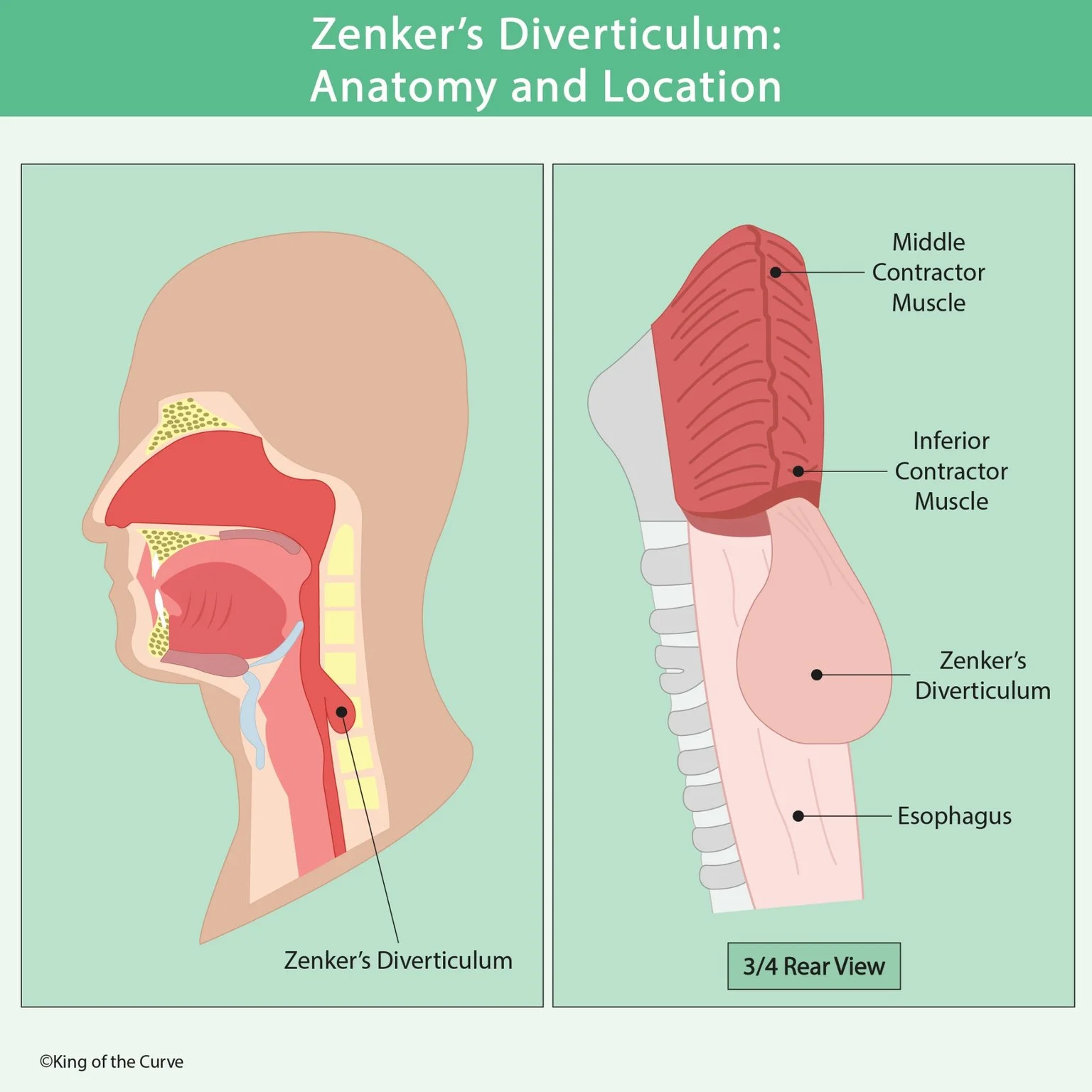
Zenker’s diverticulum is a false diverticulum, meaning that the pouch contains mucosa and submucosa but does not include all layers of the muscular wall. It develops when these inner layers protrude through a weak area of the posterior hypopharynx. Unlike a true diverticulum, which involves all layers of the organ wall, Zenker’s diverticulum is primarily associated with abnormal swallowing mechanics and increased pressure during swallowing.
📍 Where Is Zenker’s Diverticulum Located?
Zenker’s diverticulum forms at Killian’s dehiscence, a naturally weak region between the oblique fibers of the thyropharyngeus and the transverse fibers of the cricopharyngeus, both components of the inferior pharyngeal constrictor complex. This region lies immediately above the upper esophageal sphincter. The pouch usually projects posteriorly and may extend downward alongside the cervical esophagus as it enlarges.
💪 Role of the Cricopharyngeus Muscle
The cricopharyngeus muscle is a major component of the upper esophageal sphincter. Normally, it relaxes during swallowing to allow food to pass from the pharynx into the esophagus. When its relaxation or opening is impaired, pressure can increase inside the hypopharynx. Repeated pressure against Killian’s dehiscence can gradually force the mucosa and submucosa outward, forming a diverticular pouch.
🔄 How Does Zenker’s Diverticulum Develop?
The development of Zenker’s diverticulum is commonly explained as a pulsion mechanism, in which increased internal pressure pushes tissue through an area of weakness. Dysfunction of the upper esophageal sphincter contributes to this process. Over time, the pouch may enlarge and begin retaining food, saliva, and other swallowed material. This retention explains many of the characteristic symptoms, including regurgitation and unpleasant breath odor.
⚠️ Common Signs and Symptoms
Patients with Zenker’s diverticulum frequently experience progressive oropharyngeal dysphagia, particularly difficulty initiating or completing swallowing. Other symptoms include regurgitation of undigested food, halitosis, coughing, choking, and a sensation of food becoming trapped in the throat. Some patients develop a gurgling sound in the neck or experience unexplained weight loss. Regurgitated material may enter the airway, increasing the risk of aspiration and recurrent respiratory infections.
📊 Zenker’s Diverticulum vs. Other Esophageal Diverticula
Different types of diverticula can be distinguished by their anatomical location and underlying mechanism.
| Feature | Zenker's Diverticulum | Mid-Esophageal Diverticulum | Epiphrenic Diverticulum |
|---|---|---|---|
| Location | Posterior hypopharynx, above the upper esophageal sphincter | Middle esophagus | Distal esophagus, above the diaphragm |
| Typical Mechanism | Pulsion | Traditionally traction; may also be pulsion | Pulsion, often related to motility disorders |
| Wall Involvement | False diverticulum | Variable | Usually false diverticulum |
| Common Symptoms | Dysphagia, regurgitation, halitosis | Often asymptomatic; possible dysphagia | Dysphagia, regurgitation, chest discomfort |
| Major Concern | Aspiration | Depends on size and cause | Retention and aspiration |
The most important distinction is that Zenker’s diverticulum originates in the hypopharynx, whereas mid-esophageal and epiphrenic diverticula arise within the esophagus itself.
🔬 How Is Zenker’s Diverticulum Diagnosed?
A barium swallow study, also called a contrast esophagram, is commonly used to identify Zenker’s diverticulum. It demonstrates a contrast-filled pouch arising from the posterior pharyngoesophageal region. The study can reveal the diverticulum’s size, location, and relationship to the esophageal lumen. Endoscopic evaluation may also be performed when appropriate, but clinicians must exercise caution because the endoscope can inadvertently enter the pouch and cause injury.
🏥 Treatment and Potential Complications
Treatment depends on symptom severity, diverticulum size, and the patient’s overall condition. Small, asymptomatic diverticula may be observed. Symptomatic cases are often treated with endoscopic septotomy or surgical approaches that address the diverticulum and the cricopharyngeal muscle. A cricopharyngeal myotomy helps reduce the pressure contributing to pouch formation. Untreated symptomatic diverticula can lead to aspiration pneumonia, nutritional deficiencies, weight loss, and reduced quality of life.
🧠 High-Yield Anatomy and Exam Takeaways
Remember that Zenker’s diverticulum is a false pulsion diverticulum located at Killian’s dehiscence, immediately above the upper esophageal sphincter. Its formation is associated with impaired cricopharyngeal opening and increased hypopharyngeal pressure. The classic presentation includes dysphagia, regurgitation of undigested food, halitosis, and aspiration risk. A barium swallow is a key diagnostic investigation.

Frequently Asked Questions (FAQs)
-
Aim for 4-6 focused hours, ensuring you incorporate breaks to avoid burnout.
-
Practice mindfulness techniques, take practice exams under realistic conditions, and maintain a balanced lifestyle.
-
Set short-term goals, seek support from mentors, and reward yourself for small achievements.
-
Regular exercise improves focus, reduces stress, and enhances overall mental clarity.
-
KOTC offers personalized learning tools, gamification features, and adaptive question banks to help students stay on track without burnout.