
Vitamin D3 is essential for calcium absorption, phosphate balance, bone mineralization, and normal endocrine function. Unlike most vitamins, vitamin D3 can be produced in the skin through exposure to sunlight and subsequently converted into an active hormone.
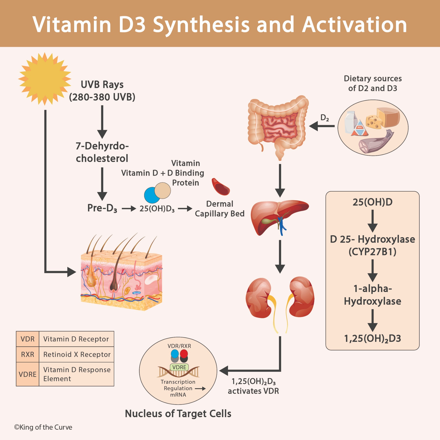
☀️ How Does Sunlight Trigger Vitamin D3 Synthesis?
Vitamin D3 synthesis begins in the epidermis, where 7-dehydrocholesterol, a cholesterol precursor, absorbs ultraviolet B (UVB) radiation.
UVB wavelengths of approximately 290–315 nm initiate a photochemical reaction that converts 7-dehydrocholesterol into previtamin D3. This molecule then undergoes a temperature-dependent rearrangement to form vitamin D3, also called cholecalciferol.
Vitamin D3 enters the circulation and is transported primarily by vitamin D-binding protein (DBP). This transport allows vitamin D3 to reach the liver for its first activation step.
MCAT Tip: UVB radiation initiates vitamin D3 synthesis, but sunlight does not directly produce the fully active hormone calcitriol.
🥗 Dietary Sources of Vitamin D2 and Vitamin D3
Vitamin D can also enter the body through food and supplements. The two major dietary forms are vitamin D2 (ergocalciferol) and vitamin D3 (cholecalciferol).
Vitamin D2 is commonly obtained from UV-exposed mushrooms and fortified foods, while vitamin D3 is found in fatty fish, egg yolks, and certain fortified products.
Both forms are absorbed in the small intestine with dietary fats, transported through the lymphatic system in chylomicrons, and eventually delivered to the liver.
Because vitamin D is fat-soluble, conditions that interfere with fat absorption can also reduce vitamin D absorption.
For the MCAT, remember that vitamin D2 and vitamin D3 are different compounds, but both can undergo hydroxylation to form biologically active vitamin D metabolites.
🩸 Vitamin D-Binding Protein: Transport Through the Blood
After vitamin D3 is synthesized in the skin, it enters the bloodstream and binds primarily to vitamin D-binding protein.
DBP transports vitamin D and its metabolites through the circulation, helping deliver them to organs involved in vitamin D metabolism.
Vitamin D obtained from food initially travels mainly in chylomicrons before entering hepatic and circulating vitamin D pools.
DBP is particularly important for transporting 25-hydroxyvitamin D and 1,25-dihydroxyvitamin D.
This illustrates an important biochemical principle: hydrophobic molecules often require carrier proteins or lipoproteins for efficient transport in aqueous biological environments.
🧬 Liver Activation: Vitamin D3 to Calcidiol
The first major hydroxylation step occurs in the liver.
The enzyme 25-hydroxylase, primarily CYP2R1, converts vitamin D3 into 25-hydroxyvitamin D3, also known as calcidiol.
Calcidiol is the major circulating form of vitamin D and is commonly measured in blood tests to evaluate vitamin D status.
Although calcidiol has some biological activity, it is not the principal active hormone responsible for vitamin D's endocrine effects.
High-yield distinction: The liver performs 25-hydroxylation, whereas the kidneys perform the major regulated 1α-hydroxylation step.
🩺 Kidney Activation: Calcidiol to Calcitriol
The second major activation step occurs primarily in the proximal tubules of the kidneys.
The enzyme 1α-hydroxylase (CYP27B1) converts calcidiol into 1,25-dihydroxyvitamin D3, also called calcitriol.
Calcitriol is the principal biologically active hormonal form of vitamin D.
Its production is regulated by parathyroid hormone (PTH), calcium balance, phosphate availability, and fibroblast growth factor 23 (FGF23).
PTH stimulates renal calcitriol synthesis, whereas FGF23 generally suppresses it.
This regulation connects vitamin D metabolism to endocrine feedback mechanisms commonly tested on the MCAT.
🧬 How Does Calcitriol Activate the Vitamin D Receptor?
Calcitriol produces many of its biological effects by binding to the intracellular vitamin D receptor (VDR), a member of the nuclear receptor superfamily.
After calcitriol binds VDR, the receptor forms a heterodimer with the retinoid X receptor (RXR).
The VDR–RXR complex interacts with specific DNA sequences called vitamin D response elements (VDREs), helping regulate transcription of target genes.
🦴 Physiological Effects of Active Vitamin D3
Calcitriol helps maintain calcium and phosphate homeostasis, primarily by increasing their absorption in the small intestine.
It regulates proteins involved in intestinal calcium transport, including calcium channels and calcium-binding proteins.
Adequate calcium and phosphate availability supports proper bone mineralization and skeletal development.
Calcitriol also participates in renal mineral handling and bone remodeling, with effects that depend on the body's physiological state and interactions with other hormones.
Vitamin D deficiency can impair bone mineralization, leading to rickets in children or osteomalacia in adults.
These effects explain why vitamin D metabolism is closely associated with bone physiology and endocrine regulation.
📊 Vitamin D3 Synthesis and Activation: High-Yield MCAT Table
The following table summarizes the major steps of vitamin D3 synthesis, transport, activation, and signaling.
| Location | Key Process | Main Product or Outcome |
|---|---|---|
| Skin | UVB acts on 7-dehydrocholesterol | Previtamin D3 → Vitamin D3 |
| Small Intestine | Absorption of dietary vitamin D2 and D3 | Vitamin D transported in chylomicrons |
| Blood | Transport by vitamin D-binding protein | Circulating vitamin D metabolites |
| Liver | 25-hydroxylation (CYP2R1) | Calcidiol — 25(OH)D |
| Kidneys | 1α-hydroxylation (CYP27B1) | Calcitriol — 1,25(OH)2D |
| Target Cells | Calcitriol binds VDR and regulates transcription | Changes in target gene expression |
🎯 MCAT High-Yield Summary: Remember the Vitamin D3 Pathway
Vitamin D3 metabolism is an excellent example of how multiple organ systems cooperate to regulate physiological function.
Remember the sequence Skin → Blood → Liver → Kidneys → Nucleus.
The skin uses UVB radiation to initiate vitamin D3 synthesis. Transport proteins carry vitamin D metabolites through the blood. The liver produces calcidiol, and the kidneys convert it into calcitriol. Finally, calcitriol binds VDR and regulates gene transcription in target cells.

Frequently Asked Questions (FAQs)
-
Aim for 4-6 focused hours, ensuring you incorporate breaks to avoid burnout.
-
Practice mindfulness techniques, take practice exams under realistic conditions, and maintain a balanced lifestyle.
-
Set short-term goals, seek support from mentors, and reward yourself for small achievements.
-
Regular exercise improves focus, reduces stress, and enhances overall mental clarity.
-
KOTC offers personalized learning tools, gamification features, and adaptive question banks to help students stay on track without burnout.