
Vitamin D is essential for calcium balance, phosphate regulation, bone mineralization, and normal physiological function. Unlike most vitamins, vitamin D can be synthesized in the skin and converted into an active hormone through a series of biochemical reactions.
☀️ How Does the Body Synthesize Vitamin D?
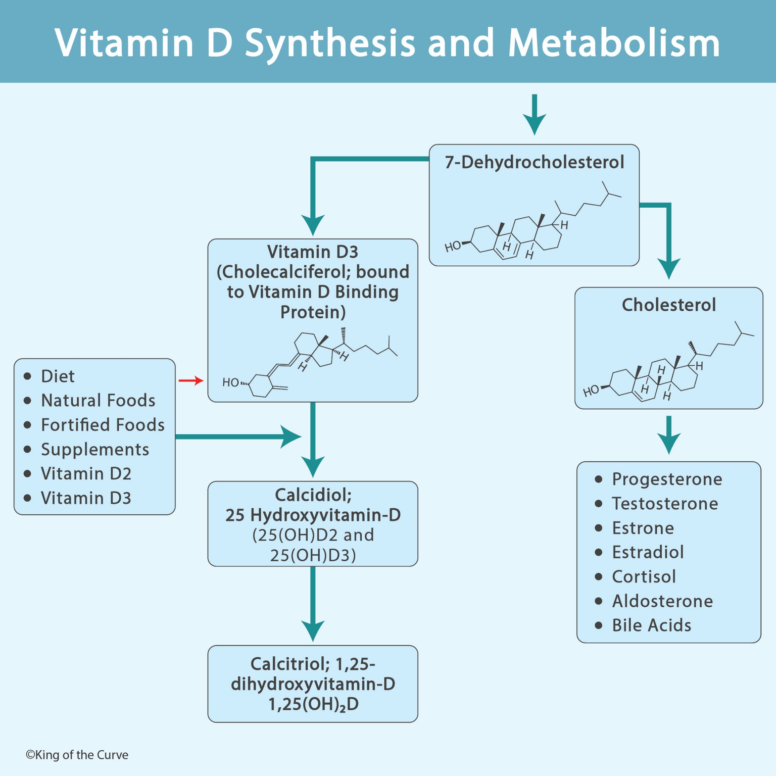
Vitamin D synthesis begins in the skin when ultraviolet B (UVB) radiation from sunlight acts on 7-dehydrocholesterol, a cholesterol-related molecule found in the epidermis.
UVB radiation converts 7-dehydrocholesterol into previtamin D3, which undergoes a heat-dependent rearrangement to form vitamin D3 (cholecalciferol).
Vitamin D3 then enters circulation, primarily bound to vitamin D-binding protein (DBP), and travels to the liver for further processing.
This pathway demonstrates how sunlight initiates the production of a molecule that ultimately functions as a hormone.
🥗 Dietary Sources of Vitamin D
Vitamin D can also be obtained from food and supplements, particularly when sunlight exposure is limited.
The two major dietary forms are:
Vitamin D2 (ergocalciferol): Derived mainly from UV-exposed fungi and yeast.
Vitamin D3 (cholecalciferol): Found in certain animal-derived foods and supplements.
Important sources include fatty fish, egg yolks, fortified milk, fortified cereals, and vitamin D supplements.
Both vitamin D2 and vitamin D3 undergo metabolic activation in the liver and kidneys. However, neither form is the fully active hormone when initially consumed.
🧬 Step 1: Vitamin D Activation in the Liver
After entering circulation, vitamin D travels to the liver, where it undergoes its first major hydroxylation reaction.
The enzyme vitamin D 25-hydroxylase, primarily CYP2R1, converts vitamin D into 25-hydroxyvitamin D, also called calcidiol.
Calcidiol is the major circulating storage and measurement form of vitamin D.
Because it has a relatively long half-life, serum 25-hydroxyvitamin D is commonly measured to assess vitamin D status.
MCAT Tip: Calcidiol is not the principal active hormonal form of vitamin D.
🩺 Step 2: Vitamin D Activation in the Kidneys
The second major activation step occurs primarily in the proximal tubules of the kidneys.
The enzyme 1α-hydroxylase (CYP27B1) converts calcidiol into calcitriol, the biologically active form of vitamin D.
Calcitriol, also known as 1,25-dihydroxyvitamin D, acts as a steroid-like hormone by binding to intracellular vitamin D receptors.
These receptors regulate gene transcription in target tissues, including the intestines, kidneys, and bones.
This is an important MCAT connection between vitamin D metabolism and steroid hormone signaling.
🦴 How Does Calcitriol Regulate Calcium and Phosphate?
Calcitriol plays a central role in maintaining calcium and phosphate homeostasis.
Its major physiological actions include increasing intestinal absorption of calcium and phosphate, supporting normal bone mineralization, and participating in the regulation of bone remodeling.
Calcitriol can also influence renal calcium handling and interact with parathyroid hormone (PTH) to maintain appropriate extracellular calcium concentrations.
When vitamin D activity is inadequate, intestinal calcium absorption decreases, potentially contributing to impaired bone mineralization.
Severe deficiency may cause rickets in children or osteomalacia in adults.
⚖️ How Is Vitamin D Metabolism Regulated?
Vitamin D activation is tightly controlled by endocrine feedback mechanisms.
When blood calcium levels fall, the parathyroid glands release parathyroid hormone (PTH). PTH stimulates renal 1α-hydroxylase, increasing calcitriol production.
Calcitriol subsequently increases intestinal calcium absorption, helping restore calcium balance.
In contrast, fibroblast growth factor 23 (FGF23), which is involved in phosphate regulation, suppresses renal calcitriol production.
Calcitriol also promotes negative feedback by limiting its own synthesis and increasing its breakdown through enzymes such as CYP24A1.
High-yield relationship: PTH generally stimulates renal calcitriol synthesis, whereas FGF23 suppresses it.
🔬 Vitamin D and Cholesterol: Understanding the Connection
Vitamin D synthesis and steroid hormone synthesis share an important biochemical connection: both involve cholesterol-related molecules.
7-Dehydrocholesterol can serve as a precursor for cholesterol synthesis or undergo UVB-dependent conversion toward vitamin D3 production.
Cholesterol, meanwhile, serves as the precursor for several steroid hormones and bile acids.
| Cholesterol-Related Pathway | Major Products | Main Function |
|---|---|---|
| Vitamin D Pathway | Cholecalciferol → Calcidiol → Calcitriol | Calcium and phosphate homeostasis |
| Glucocorticoid Synthesis | Cortisol | Stress response and metabolism |
| Mineralocorticoid Synthesis | Aldosterone | Sodium, potassium, and blood pressure regulation |
| Sex Steroid Synthesis | Progesterone, testosterone, estradiol, estrone | Reproduction and sexual development |
| Bile Acid Synthesis | Primary bile acids | Fat digestion and absorption |
🧠 Vitamin D Deficiency and Clinical MCAT Connections
Vitamin D deficiency may result from inadequate dietary intake, limited sunlight exposure, intestinal malabsorption, liver disease, or impaired kidney activation.
Chronic kidney disease is especially important because reduced renal 1α-hydroxylase activity can decrease calcitriol production.
Reduced calcitriol contributes to impaired intestinal calcium absorption and can promote secondary hyperparathyroidism.
For the MCAT, remember these associations: vitamin D deficiency can impair bone mineralization; liver dysfunction may interfere with 25-hydroxylation; and kidney dysfunction can reduce the production of active calcitriol.
Also remember that 25-hydroxyvitamin D, rather than calcitriol, is the standard laboratory marker used to evaluate vitamin D stores.

Frequently Asked Questions (FAQs)
-
Aim for 4-6 focused hours, ensuring you incorporate breaks to avoid burnout.
-
Practice mindfulness techniques, take practice exams under realistic conditions, and maintain a balanced lifestyle.
-
Set short-term goals, seek support from mentors, and reward yourself for small achievements.
-
Regular exercise improves focus, reduces stress, and enhances overall mental clarity.
-
KOTC offers personalized learning tools, gamification features, and adaptive question banks to help students stay on track without burnout.