
🚑 Ureteropelvic Junction Obstruction and Hydronephrosis
The ureteropelvic junction (UPJ) is the point where the renal pelvis transitions into the ureter, allowing urine to flow from the kidney to the bladder. When this junction becomes narrowed or obstructed, urine cannot drain properly, leading to a condition known as hydronephrosis.
🔍 What Is Ureteropelvic Junction Obstruction?
Ureteropelvic junction obstruction occurs when urine flow is partially or completely blocked at the junction between the kidney and the ureter. This obstruction causes urine to back up into the kidney, resulting in progressive dilation of the renal pelvis and calyces.
UPJ obstruction may be:
Congenital (most common, due to abnormal muscle development or crossing vessels)
Acquired (from kidney stones, scarring, infection, or trauma)
💧 How Does Hydronephrosis Develop?
When urine cannot pass efficiently through the UPJ:
Urine accumulates in the renal pelvis
The pelvis and calyces become dilated
Persistent pressure damages renal tissue
Kidney function may decline over time
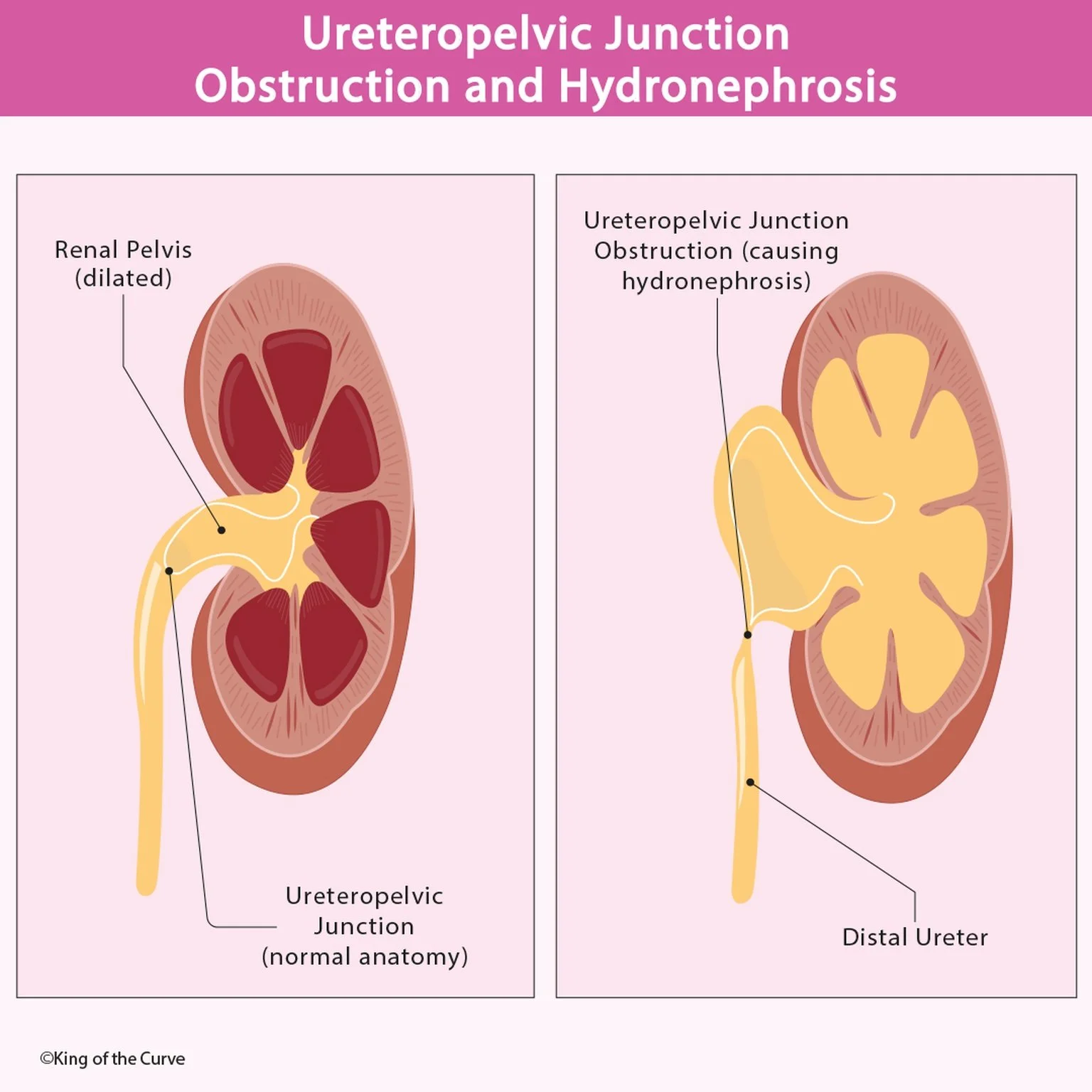
In the infographic:
The left image shows normal anatomy with proper urine drainage.
The right image demonstrates obstruction at the UPJ, leading to marked dilation and hydronephrosis.
📊 Ureteropelvic Junction Obstruction vs Normal Anatomy
| Feature | Normal Ureteropelvic Junction | UPJ Obstruction with Hydronephrosis |
|---|---|---|
| Urine flow | Free flow from renal pelvis to ureter | Partially or completely obstructed |
| Renal pelvis | Normal size | Dilated |
| Calyces | Normal shape | Dilated and blunted |
| Kidney pressure | Normal intrarenal pressure | Increased pressure within kidney |
| Renal parenchyma | Preserved | Progressive thinning with chronic obstruction |
| Symptoms | Usually asymptomatic | Flank pain, UTIs, hematuria, nausea |
| Risk to kidney function | None | Risk of progressive renal damage |
| Management | No treatment needed | Observation or surgical correction |
⚠️ Clinical Features
Symptoms vary with age and severity:
Infants: Antenatal hydronephrosis on ultrasound
Children: Abdominal mass, urinary tract infections
Adults: Flank pain, nausea, hematuria, recurrent UTIs
Chronic obstruction can lead to irreversible renal damage if untreated.
🧪 Diagnosis
UPJ obstruction is diagnosed using:
Ultrasound (detects hydronephrosis)
CT urography
Diuretic renography (assesses drainage and renal function)
🛠️ Management
Treatment depends on severity:
Observation for mild, asymptomatic cases
Surgical correction (pyeloplasty) for significant obstruction
Endoscopic or minimally invasive approaches in select patients
Early intervention helps preserve kidney function and prevent complications.
✅ Key Takeaway
Ureteropelvic junction obstruction is a common cause of hydronephrosis, especially in children. Recognizing the anatomical blockage and understanding its effect on urine flow is essential for timely diagnosis and management.

Frequently Asked Questions (FAQs)
-
Aim for 4-6 focused hours, ensuring you incorporate breaks to avoid burnout.
-
Practice mindfulness techniques, take practice exams under realistic conditions, and maintain a balanced lifestyle.
-
Set short-term goals, seek support from mentors, and reward yourself for small achievements.
-
Regular exercise improves focus, reduces stress, and enhances overall mental clarity.
-
KOTC offers personalized learning tools, gamification features, and adaptive question banks to help students stay on track without burnout.