
💧 Tubular Fluid Osmolarity in Low ADH: Understanding the Nephron
Antidiuretic hormone (ADH), also called vasopressin, plays an important role in regulating the body’s water balance. Its major renal effect is to increase water permeability in the collecting ducts, allowing more water to be reabsorbed into the bloodstream. When ADH levels are low, the collecting ducts become relatively impermeable to water, resulting in the production of more dilute urine.
🩸 Starting in the Proximal Tubule
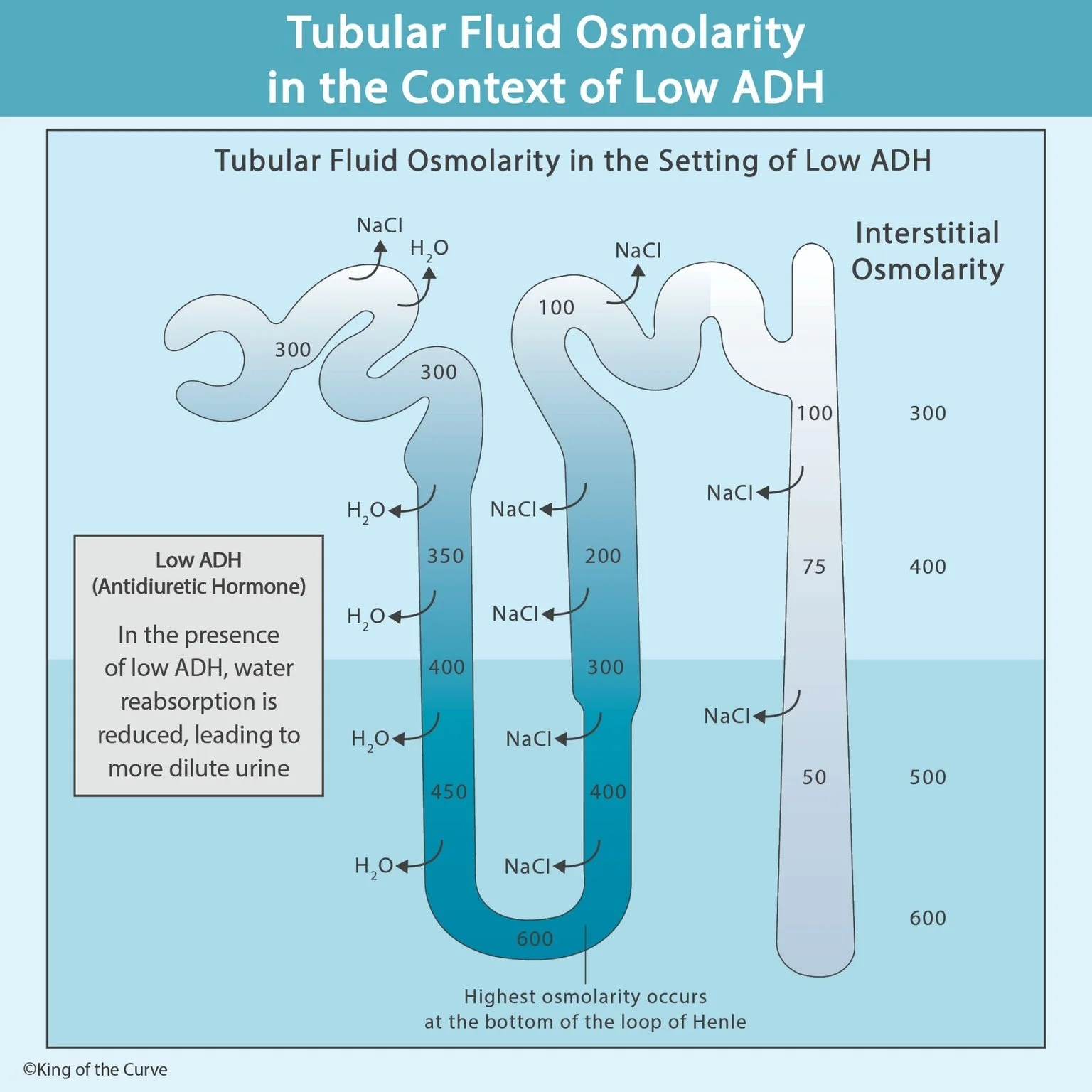
Tubular fluid entering the nephron is approximately isotonic with plasma. In the proximal convoluted tubule, water and solutes such as sodium are reabsorbed together. Because their removal is roughly proportional, tubular fluid osmolarity remains near 300 mOsm/L through much of this segment.
⬇️ Descending Loop of Henle
The descending limb is highly permeable to water but much less permeable to NaCl. As the tubule travels deeper into the increasingly hyperosmotic renal medulla, water leaves the tubular fluid. This causes the fluid inside the nephron to become progressively more concentrated.
🔄 The Bottom of the Loop
Tubular fluid reaches its greatest concentration near the bottom of the loop of Henle. The exact osmolarity varies with hydration state and the strength of the medullary osmotic gradient. The key MCAT concept is the direction of change: osmolarity rises in the descending limb and then falls in the ascending limb.
⬆️ Ascending Loop of Henle
The ascending limb behaves very differently. It is essentially impermeable to water while NaCl leaves the tubular fluid. In the thick ascending limb, Na⁺, K⁺, and Cl⁻ are actively reabsorbed through the NKCC2 cotransporter. Because salt leaves without water following it, the tubular fluid becomes progressively more dilute.
🧪 The Diluting Segment
By the time tubular fluid reaches the distal nephron, its osmolarity may be substantially below that of plasma. This is why the thick ascending limb and early distal tubule are often described as diluting segments. They remove solute while retaining water within the tubular fluid.
| 🧬 Nephron Region | 💧 Water Movement | 🧂 Major Solute Effect | 📊 Tubular Fluid |
|---|---|---|---|
| Proximal tubule | Water reabsorbed | Solutes reabsorbed | ~ Isotonic |
| Descending limb | Water exits | Little NaCl removal | More concentrated |
| Ascending limb | Water cannot follow | NaCl reabsorbed | More dilute |
| Collecting duct, low ADH | Little water reabsorbed | Variable solute handling | Very dilute |
🚰 Collecting Duct During Low ADH
Low ADH means fewer aquaporin-2 water channels are inserted into the apical membrane of collecting-duct principal cells. Even though the surrounding medullary interstitium becomes increasingly concentrated deeper in the kidney, relatively little water can leave the collecting duct. The tubular fluid therefore remains dilute.
⚖️ Why Low ADH Produces Dilute Urine
The kidney has already removed substantial amounts of NaCl from the tubular fluid before it reaches the collecting duct. With low ADH, the collecting duct cannot efficiently recover water in response to the medullary osmotic gradient. Consequently, a larger volume of relatively dilute urine is excreted.
🎯 MCAT High-Yield Takeaway
Remember the pattern: descending limb = water leaves; ascending limb = salt leaves; low ADH = less water leaves the collecting duct. ADH does not create the medullary gradient itself; rather, it allows the collecting duct to take advantage of that gradient by increasing water permeability. This relationship between nephron permeability, osmolarity, and hormonal regulation is a high-yield MCAT renal physiology concept.

Frequently Asked Questions (FAQs)
-
Aim for 4-6 focused hours, ensuring you incorporate breaks to avoid burnout.
-
Practice mindfulness techniques, take practice exams under realistic conditions, and maintain a balanced lifestyle.
-
Set short-term goals, seek support from mentors, and reward yourself for small achievements.
-
Regular exercise improves focus, reduces stress, and enhances overall mental clarity.
-
KOTC offers personalized learning tools, gamification features, and adaptive question banks to help students stay on track without burnout.