
🫁 Thoracic Cavity and Mediastinum: Anatomy Made Simple
The thoracic cavity is the region of the body enclosed by the rib cage, sternum, thoracic vertebrae, and diaphragm. It contains essential structures involved in respiration, circulation, digestion, and nervous system function. For anatomy and MCAT preparation, understanding how this cavity is divided helps organize many structures that can otherwise seem difficult to memorize.
🫀 What Is the Mediastinum?
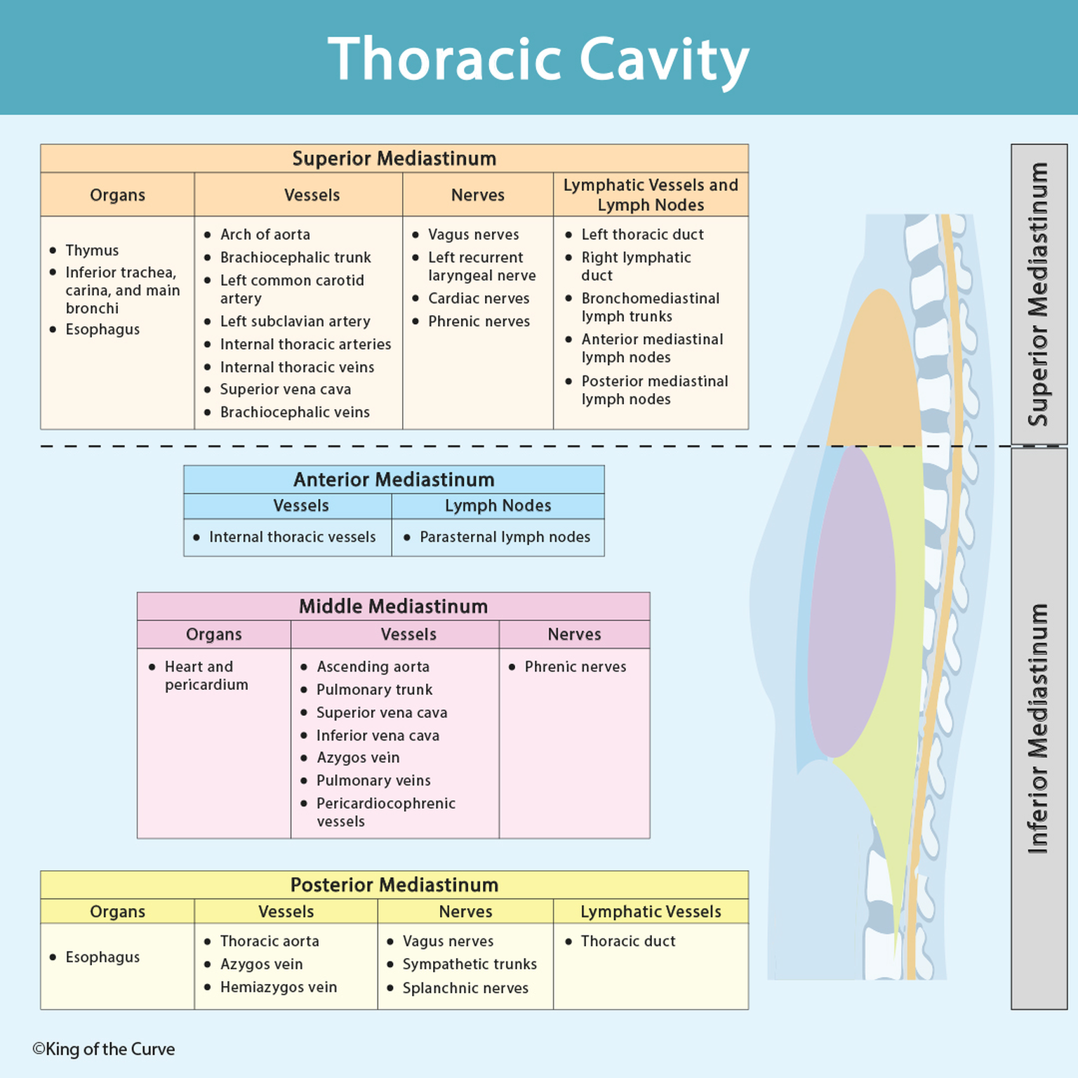
The mediastinum is the central compartment of the thoracic cavity located between the two pleural cavities. It contains the heart, major blood vessels, trachea, esophagus, thymus, nerves, and lymphatic structures. Anatomically, it is divided into the superior mediastinum and inferior mediastinum, with the inferior portion further divided into anterior, middle, and posterior compartments.
🔝 Superior Mediastinum
The superior mediastinum lies above the transverse thoracic plane. Important structures found here include the thymus, trachea, esophagus, aortic arch and its major branches, superior vena cava, vagus nerves, phrenic nerves, and recurrent laryngeal nerve. The thoracic duct and several groups of mediastinal lymph nodes also pass through this region.
🔵 Anterior Mediastinum
The anterior mediastinum is the relatively small space between the sternum and pericardium within the inferior mediastinum. It contains loose connective tissue, fat, lymphatic structures, and remnants of the thymus in adults. Internal thoracic vessels and parasternal lymph nodes are closely associated with this anterior thoracic region.
❤️ Middle Mediastinum
The middle mediastinum contains the heart and pericardium, making it one of the most clinically important thoracic compartments. It also contains structures associated with the heart, including the ascending aorta, pulmonary trunk, pulmonary veins, superior and inferior venae cavae, and phrenic nerves.
🟡 Posterior Mediastinum
The posterior mediastinum lies behind the pericardium and in front of the thoracic vertebral column. Major structures include the descending thoracic aorta, esophagus, thoracic duct, azygos and hemiazygos veins, vagus nerves, sympathetic trunks, and thoracic splanchnic nerves.
🧠 Key Mediastinal Compartments
| 🫁 Compartment | 📘 Important Structures |
|---|---|
| Superior | Thymus, trachea, esophagus, aortic arch, superior vena cava, vagus nerves, and phrenic nerves |
| Anterior | Fat, connective tissue, thymic remnants, and lymphatic structures |
| Middle | Heart, pericardium, great vessels, and phrenic nerves |
| Posterior | Esophagus, thoracic aorta, thoracic duct, azygos system, vagus nerves, and splanchnic nerves |
🩸 Why These Divisions Matter
Dividing the mediastinum into compartments makes it easier to predict where anatomical structures, masses, or abnormalities may be located. The relationships are also useful for understanding cardiovascular, respiratory, gastrointestinal, lymphatic, and autonomic pathways through the chest.
🎯 MCAT High-Yield Takeaway
For the MCAT, focus on the big anatomical relationships rather than memorizing every small structure. Remember: superior mediastinum = major passageway structures, middle mediastinum = heart and pericardium, and posterior mediastinum = esophagus, descending aorta, thoracic duct, and autonomic nerves. Connecting each compartment with its major structures makes thoracic anatomy much easier to recall.

Frequently Asked Questions (FAQs)
-
Aim for 4-6 focused hours, ensuring you incorporate breaks to avoid burnout.
-
Practice mindfulness techniques, take practice exams under realistic conditions, and maintain a balanced lifestyle.
-
Set short-term goals, seek support from mentors, and reward yourself for small achievements.
-
Regular exercise improves focus, reduces stress, and enhances overall mental clarity.
-
KOTC offers personalized learning tools, gamification features, and adaptive question banks to help students stay on track without burnout.