
🩸 Schistocytes: What Are Helmet Cells and Why Do They Matter?
Schistocytes are fragmented red blood cells (RBCs) seen on a peripheral blood smear. They are formed when erythrocytes are mechanically damaged while circulating in the bloodstream. Because of their irregular, sharp-edged appearance, schistocytes are also known as “helmet cells.”
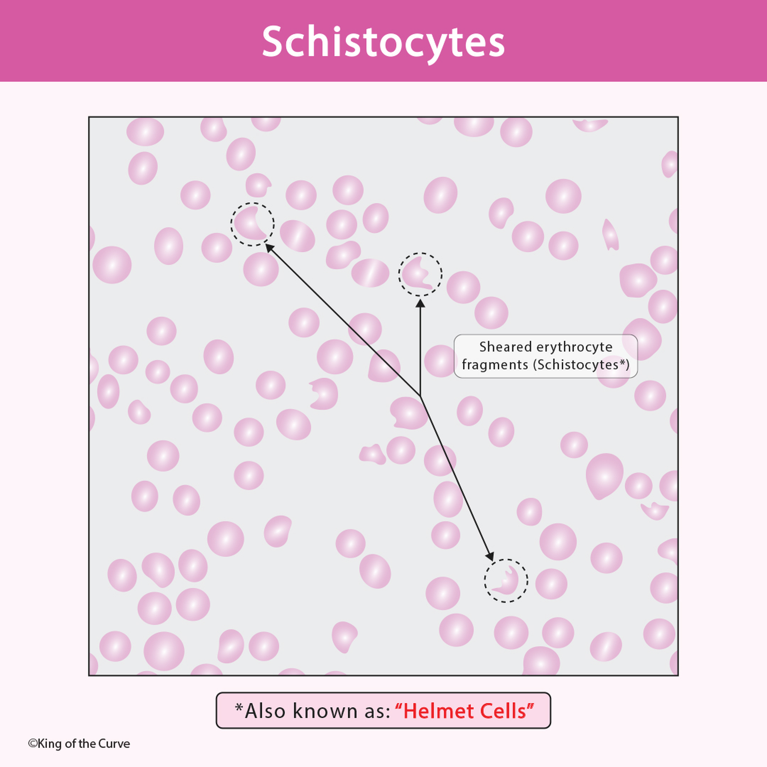
🔬 What Are Schistocytes?
Schistocytes are sheared erythrocyte fragments produced when red blood cells are physically torn apart. Instead of appearing as smooth, round biconcave discs, they look:
Irregularly shaped
Crescent-shaped
Triangular or helmet-like
Jagged with sharp edges
These fragments result from mechanical destruction within the circulation.
⚙️ How Do Schistocytes Form?
Schistocytes typically form when red blood cells pass through:
Narrowed or obstructed blood vessels
Fibrin strands within small vessels
Artificial heart valves
In these situations, RBCs are forced through abnormal structures, leading to mechanical shearing and fragmentation.
This process is called intravascular hemolysis.
🚨 Clinical Significance
The presence of schistocytes on a peripheral smear is highly suggestive of microangiopathic hemolytic anemia (MAHA).
🧠 Common Conditions Associated with Schistocytes
Thrombotic Thrombocytopenic Purpura (TTP)
Hemolytic Uremic Syndrome (HUS)
Disseminated Intravascular Coagulation (DIC)
HELLP syndrome
Mechanical heart valves
Severe hypertension
In these disorders, small clots or fibrin strands damage circulating RBCs.
🧪 Laboratory Findings
When schistocytes are present, labs often show:
Elevated LDH
Elevated indirect bilirubin
Decreased haptoglobin
Reticulocytosis
Anemia
Possible thrombocytopenia
These findings reflect ongoing red blood cell destruction.
🔎 Schistocytes vs Other Abnormal RBC Shapes
It is important not to confuse schistocytes with:
Spherocytes
Target cells
Sickle cells
Burr cells (echinocytes)
Schistocytes are fragmented and irregular, not uniformly shaped.
🛡 Why Are They Called Helmet Cells?
The term “helmet cell” comes from their characteristic shape. Many schistocytes resemble:
A helmet
A bite taken out of a cell
A crescent with sharp edges
This nickname helps students visually remember their appearance on smear.
🎯 Exam Tip (USMLE / MCAT / Nursing Exams)
If you see:
Anemia
Thrombocytopenia
Elevated LDH
Fragmented RBCs
Think: Microangiopathic Hemolytic Anemia
And if schistocytes are present, immediately consider:
TTP, HUS, or DIC
📌 Key Takeaway
Schistocytes are fragmented red blood cells caused by mechanical destruction in the bloodstream. Their presence on a peripheral smear is a critical diagnostic clue for life-threatening conditions such as TTP, HUS, and DIC.
Recognizing them quickly can guide urgent and potentially life-saving treatment.

Frequently Asked Questions (FAQs)
-
Aim for 4-6 focused hours, ensuring you incorporate breaks to avoid burnout.
-
Practice mindfulness techniques, take practice exams under realistic conditions, and maintain a balanced lifestyle.
-
Set short-term goals, seek support from mentors, and reward yourself for small achievements.
-
Regular exercise improves focus, reduces stress, and enhances overall mental clarity.
-
KOTC offers personalized learning tools, gamification features, and adaptive question banks to help students stay on track without burnout.