
Lymph nodes are small, bean-shaped organs of the lymphatic system that play an essential role in immune defense. They filter lymphatic fluid, trap foreign particles, and provide an environment where immune cells recognize and respond to pathogens.
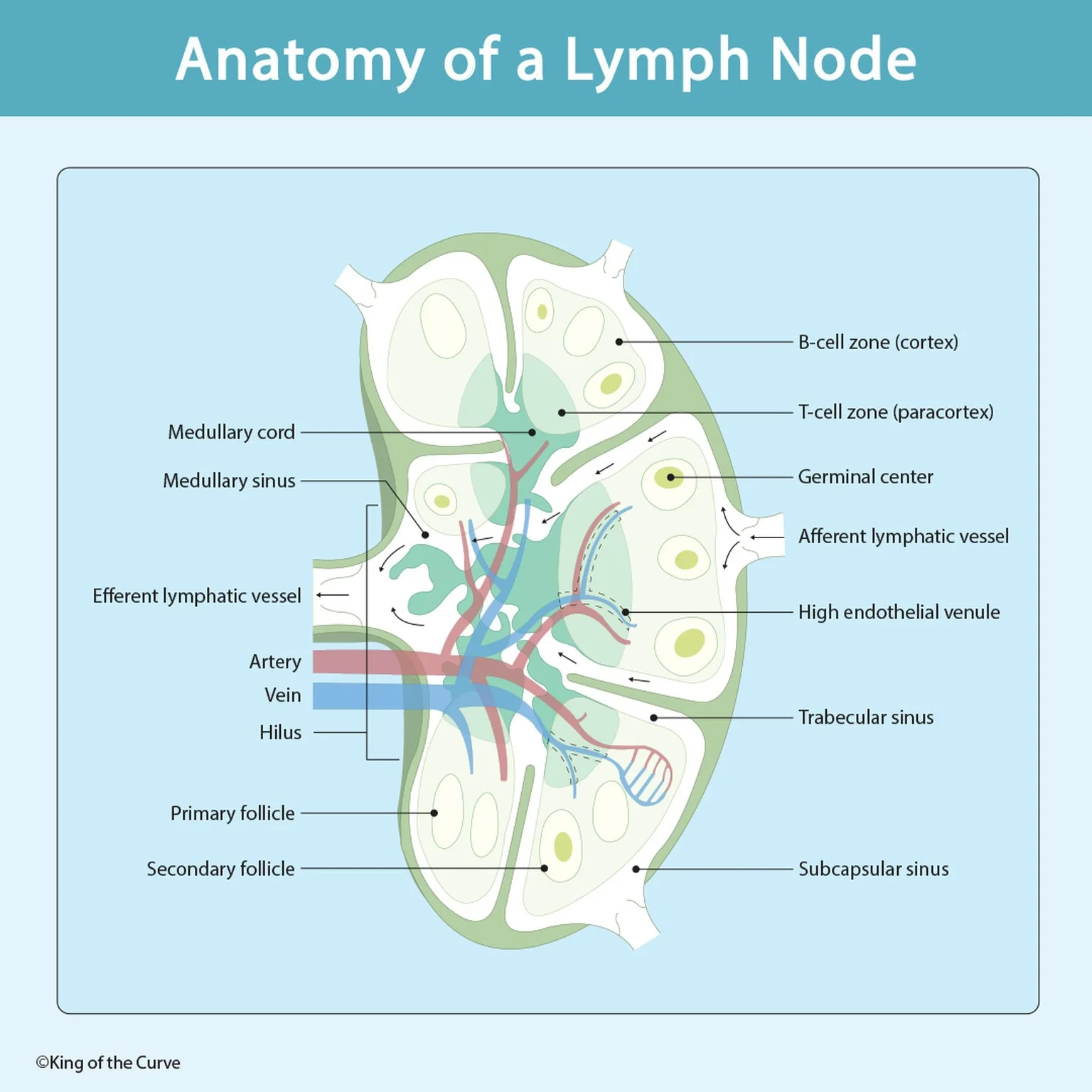
🔬 What Is a Lymph Node?
A lymph node is a secondary lymphoid organ that filters lymph and supports immune surveillance. Lymph nodes are distributed throughout the body, with prominent groups in the cervical, axillary, and inguinal regions.
Lymph enters through afferent lymphatic vessels, travels through specialized sinuses, and exits through efferent lymphatic vessels at the hilum.
As lymph passes through the node, immune cells encounter antigens, helping initiate adaptive immune responses.
MCAT Tip: Lymph nodes filter lymph, whereas the spleen primarily filters blood.
🧫 Cortex: The B-Cell Zone
The outer region of a lymph node is called the cortex. It contains lymphoid follicles populated primarily by B lymphocytes.
Two important follicle types are found here:
Primary follicles: Contain predominantly naïve B cells and lack a prominent germinal center.
Secondary follicles: Contain germinal centers that develop following antigen-dependent B-cell activation.
Within germinal centers, activated B cells proliferate and undergo processes such as somatic hypermutation and affinity maturation. Class-switch recombination can also occur during the germinal-center response.
These processes help generate effective antibody responses and immunological memory.
High-yield distinction: The cortex is primarily associated with B cells and antibody-mediated immunity.
🛡️ Paracortex: The T-Cell Zone
The paracortex lies between the cortex and medulla and contains large populations of T lymphocytes.
Dendritic cells present processed antigens to T cells using major histocompatibility complex (MHC) molecules.
CD4⁺ helper T cells recognize peptides presented on MHC class II molecules, while CD8⁺ cytotoxic T cells recognize peptides presented on MHC class I molecules.
The paracortex also contains high endothelial venules (HEVs), specialized blood vessels that allow circulating naïve lymphocytes to enter lymph nodes from the bloodstream.
This makes the paracortex important for T-cell activation and lymphocyte trafficking.
🩸 Medulla: Medullary Cords and Sinuses
The medulla forms the inner region of the lymph node and contains medullary cords and medullary sinuses.
Medullary cords contain plasma cells, B cells, macrophages, and other immune cells. Plasma cells produce antibodies following B-cell differentiation.
Medullary sinuses are lymph-filled channels containing macrophages and other cells involved in filtering lymph.
As lymph moves through these channels, macrophages can capture and remove microorganisms, cellular debris, and foreign material.
For the MCAT, remember that the medulla supports both lymph filtration and antibody production.
💧 Afferent vs. Efferent Lymphatic Vessels
Lymphatic vessels regulate fluid movement through lymph nodes.
Afferent lymphatic vessels enter the node along its convex surface, delivering lymph that may contain antigens and antigen-presenting cells.
Efferent lymphatic vessels leave through the hilum, carrying filtered lymph and immune cells away from the node.
The general pathway is:
Afferent vessels → Subcapsular sinus → Trabecular (cortical) sinuses → Medullary sinuses → Efferent vessel
This organized flow allows immune cells to examine lymph for potential threats.
MCAT Tip: Afferent means entering the lymph node; efferent means exiting it.
🧬 Germinal Centers and Antibody Production
Germinal centers are specialized structures that develop within secondary lymphoid follicles during many T-dependent immune responses.
Activated B cells undergo rapid proliferation and selection for improved antigen binding.
T follicular helper cells and follicular dendritic cells support this process.
Selected B cells can differentiate into antibody-secreting plasma cells or memory B cells.
This explains how the adaptive immune system produces increasingly effective antibodies after antigen exposure.
Germinal-center reactions are especially important for understanding vaccination, immunological memory, and humoral immunity.
🫀 Hilum, Blood Vessels, and Lymphocyte Circulation
The hilum is the indented region of a lymph node where the efferent lymphatic vessel exits and blood vessels enter or leave.
Arteries supply oxygenated blood and nutrients, while veins drain blood from the organ.
Lymphocytes circulate continuously between the blood, lymphatic system, and secondary lymphoid organs.
Many naïve lymphocytes enter lymph nodes through high endothelial venules, while activated and recirculating lymphocytes can leave through efferent lymphatic vessels.
This circulation increases the likelihood that rare antigen-specific lymphocytes will encounter their corresponding antigens.
📊 Lymph Node Anatomy: High-Yield MCAT Comparison Table
The following table summarizes the major anatomical structures and their immune functions.
| Structure | Location | Main Function |
|---|---|---|
| Cortex | Outer region | Contains B-cell follicles |
| Primary follicle | Cortex | Houses predominantly naïve B cells |
| Secondary follicle | Cortex | Supports activated B-cell responses |
| Germinal center | Secondary follicle | B-cell proliferation, affinity maturation, and selection |
| Paracortex | Between cortex and medulla | T-cell activation and antigen presentation |
| High endothelial venule | Primarily paracortex | Allows lymphocytes to enter from blood |
| Medullary cords | Inner region | Contain plasma cells and other immune cells |
| Medullary sinuses | Medulla | Filter lymph through macrophage-rich channels |
| Afferent lymphatic vessels | Convex surface | Carry lymph into the node |
| Efferent lymphatic vessel | Hilum | Carries lymph out of the node |
| Hilum | Indented surface | Passage for blood vessels and efferent lymphatics |
🎯 MCAT High-Yield Summary: Remember Lymph Node Anatomy
Lymph node anatomy demonstrates how the immune system organizes different cell types to detect pathogens and coordinate adaptive immunity.
Remember these essential relationships:
Cortex = B cells
Paracortex = T cells
Medullary cords = Plasma cells
Medullary sinuses = Lymph filtration
HEVs = Lymphocyte entry from blood
Afferent vessels = Lymph enters
Efferent vessels = Lymph exits

Frequently Asked Questions (FAQs)
-
Aim for 4-6 focused hours, ensuring you incorporate breaks to avoid burnout.
-
Practice mindfulness techniques, take practice exams under realistic conditions, and maintain a balanced lifestyle.
-
Set short-term goals, seek support from mentors, and reward yourself for small achievements.
-
Regular exercise improves focus, reduces stress, and enhances overall mental clarity.
-
KOTC offers personalized learning tools, gamification features, and adaptive question banks to help students stay on track without burnout.