
🫀 Placement of ECG Electrodes
The Electrocardiogram (ECG or EKG) is a vital diagnostic tool used to measure the electrical activity of the heart. Proper placement of ECG electrodes ensures accurate readings and helps detect arrhythmias, myocardial infarction, and other cardiac abnormalities. Misplacement, on the other hand, can lead to distorted waveforms and incorrect interpretations.
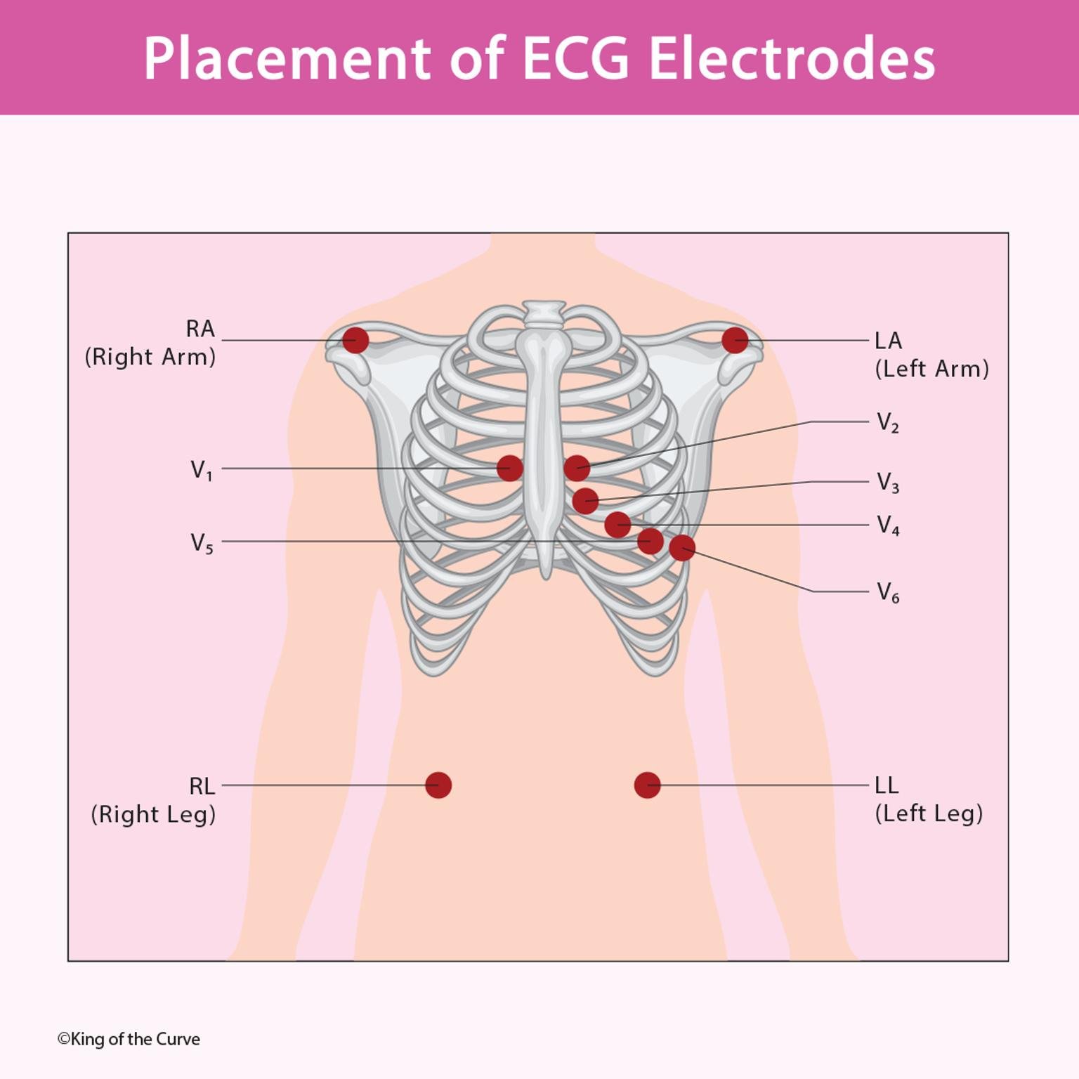
⚡ Understanding ECG Leads
An ECG records the heart’s electrical signals using 10 electrodes that produce 12 leads—six limb leads and six precordial (chest) leads. Each lead views the heart’s activity from a different angle, providing a comprehensive picture of cardiac function.
🦴 Chest (Precordial) Electrode Placement
| Lead | Placement Location |
|---|---|
| V1 | 4th intercostal space, right sternal border |
| V2 | 4th intercostal space, left sternal border |
| V3 | Midway between V2 and V4 |
| V4 | 5th intercostal space, midclavicular line |
| V5 | Level with V4, anterior axillary line |
| V6 | Level with V5, midaxillary line |
Each of these electrodes provides a “horizontal” view of the heart, particularly useful for analyzing the anterior and lateral walls of the left ventricle.
💪 Limb Electrode Placement
| Lead | Placement Location |
|---|---|
| RA (Right Arm) | Anywhere between right shoulder and wrist |
| LA (Left Arm) | Anywhere between left shoulder and wrist |
| RL (Right Leg) | Anywhere below the right torso, serves as ground |
| LL (Left Leg) | Anywhere below the left torso |
These electrodes measure electrical activity in the frontal plane, forming the basis for leads I, II, III, aVR, aVL, and aVF.
💡 Importance of Proper Electrode Placement
Correct placement is essential for reliable ECG interpretation. Incorrectly placed leads can cause:
Inverted or flattened waveforms
Pseudo-pathological findings (false positives for ischemia or infarction)
Misdiagnosis of arrhythmias or conduction blocks
Healthcare professionals must follow standardized placement guidelines to ensure consistency and diagnostic accuracy.
🩺 Clinical Significance
An ECG is not only used for diagnosing heart disease but also for monitoring patient status during surgery, anesthesia, and critical care. Accurate electrode placement enables clinicians to identify:
Atrial and ventricular hypertrophy
Ischemic changes
Conduction abnormalities
Drug-induced cardiac effects
🧠 Advanced ECG Interpretation Skills
As technology evolves, ECG machines now offer digital integration, allowing for more precise analysis and real-time monitoring. However, even the most advanced systems rely heavily on correct electrode positioning to generate meaningful data. Mastering this skill remains fundamental for all healthcare practitioners involved in cardiac care.

Frequently Asked Questions (FAQs)
-
Aim for 4-6 focused hours, ensuring you incorporate breaks to avoid burnout.
-
Practice mindfulness techniques, take practice exams under realistic conditions, and maintain a balanced lifestyle.
-
Set short-term goals, seek support from mentors, and reward yourself for small achievements.
-
Regular exercise improves focus, reduces stress, and enhances overall mental clarity.
-
KOTC offers personalized learning tools, gamification features, and adaptive question banks to help students stay on track without burnout.