
🧠 PET Scan Process on a Molecular Level
A Positron Emission Tomography (PET) scan is a powerful imaging technique that provides deep insights into the molecular and metabolic functions of tissues and organs. Unlike traditional scans that show structure, PET scans visualize how the body’s cells are functioning, making it an essential tool for diagnosing cancer, neurological disorders, and cardiovascular diseases.
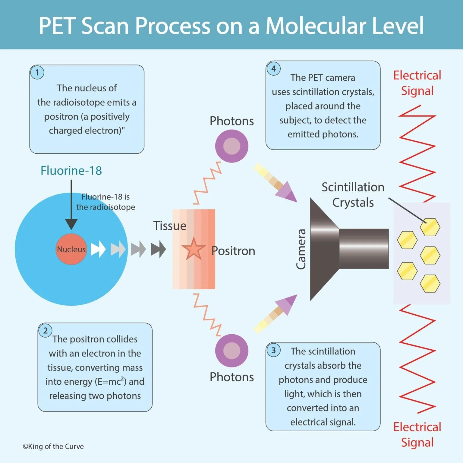
⚛️ Step 1: Emission of the Positron
The process begins with the radioisotope, such as Fluorine-18, which is incorporated into biologically active molecules like glucose. When introduced into the body, the nucleus of the radioisotope emits a positron — a positively charged electron. This emission marks the start of the molecular-level imaging process.
💥 Step 2: Annihilation Reaction
As the positron travels through the body’s tissue, it eventually collides with an electron. This collision causes a mutual annihilation event, converting their combined mass into energy according to Einstein’s equation, E = mc². The result is the release of two high-energy photons, which travel in opposite directions.
💡 Step 3: Photon Detection by Scintillation Crystals
The emitted photons reach scintillation crystals located in the PET camera. These crystals absorb the photon energy and emit flashes of light (scintillations). Each flash represents an annihilation event, capturing vital information about where the radioisotope was located in the body.
🔌 Step 4: Conversion to Electrical Signals
The scintillation light is then detected by photodetectors, which convert it into electrical signals. These electrical signals are processed by the computer system to reconstruct a detailed 3D image that shows the concentration and activity of the tracer within different tissues.
🧬 Clinical Significance
The resulting PET images allow physicians to:
Detect cancerous growths based on abnormal glucose uptake.
Evaluate brain activity in neurological conditions like Alzheimer’s or epilepsy.
Assess heart function and detect ischemic damage.
By capturing cellular metabolism in real-time, PET scans have transformed diagnostic medicine and continue to enhance personalized treatment strategies.
📊 Summary Table
| Step | Process | Key Component | Result |
|---|---|---|---|
| 1 | Emission of positron | Fluorine-18 radioisotope | Positron released |
| 2 | Annihilation reaction | Electron–positron collision | Two photons emitted |
| 3 | Photon absorption | Scintillation crystals | Light production |
| 4 | Signal conversion | Photodetector and camera | Electrical signal and image reconstruction |

Frequently Asked Questions (FAQs)
-
Aim for 4-6 focused hours, ensuring you incorporate breaks to avoid burnout.
-
Practice mindfulness techniques, take practice exams under realistic conditions, and maintain a balanced lifestyle.
-
Set short-term goals, seek support from mentors, and reward yourself for small achievements.
-
Regular exercise improves focus, reduces stress, and enhances overall mental clarity.
-
KOTC offers personalized learning tools, gamification features, and adaptive question banks to help students stay on track without burnout.