
The auditory pathway is responsible for transmitting sound information from the inner ear to the cerebral cortex, where sound is consciously perceived and interpreted. This pathway involves several important structures, including the cochlear nuclei, superior olivary complex, lateral lemniscus, inferior colliculus, and medial geniculate body.
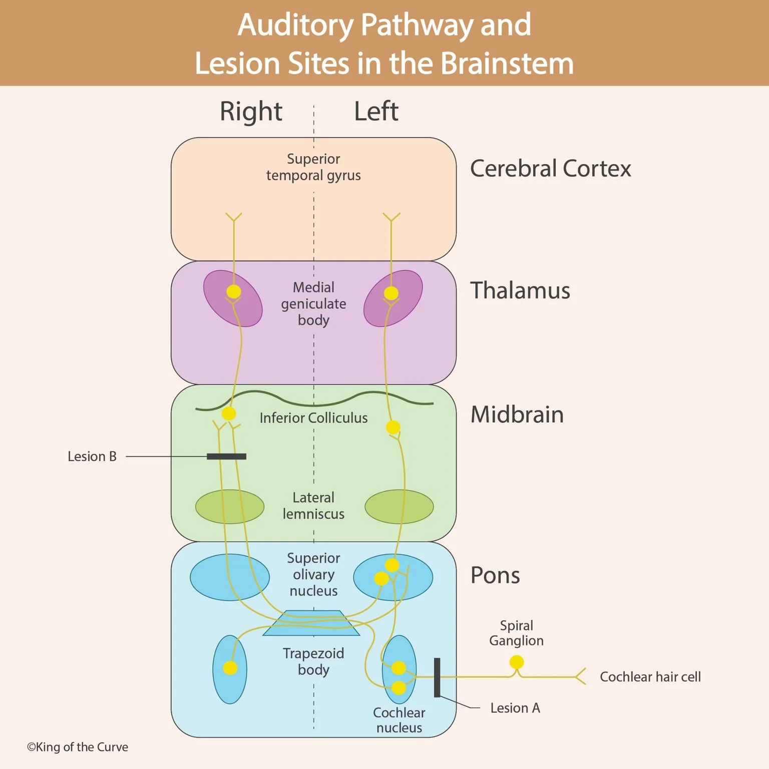
👂 How Does the Auditory Pathway Begin?
Hearing begins when sound waves enter the external auditory canal and cause the tympanic membrane to vibrate.
These vibrations travel through the middle-ear ossicles—the malleus, incus, and stapes—before reaching the oval window of the cochlea.
Within the cochlea, mechanical vibrations generate fluid movement that displaces the basilar membrane. This movement bends the stereocilia of hair cells in the organ of Corti.
Hair-cell depolarization leads to neurotransmitter release onto sensory neurons whose cell bodies are located in the spiral ganglion.
The central processes of these neurons form the cochlear division of the vestibulocochlear nerve (cranial nerve VIII), which carries auditory information toward the brainstem.
MCAT Tip: Hair cells are mechanoreceptors that convert mechanical vibrations into electrical signals through mechanotransduction.
🧬 Cochlear Nuclei: The First Brainstem Relay
Auditory information from the cochlear nerve enters the brainstem near the junction of the pons and medulla.
The nerve fibers synapse in the dorsal and ventral cochlear nuclei, which represent the first major central processing stations of the auditory pathway.
These nuclei process different aspects of sound, including timing, intensity, and frequency-related information.
Importantly, each cochlear nerve initially carries information from the ear on the same side of the body.
Damage to the cochlear nerve or cochlear nuclei can therefore produce substantial hearing impairment on the affected side.
This differs from lesions higher in the auditory pathway, where information from both ears is represented.
🔄 Superior Olivary Complex: Bilateral Sound Processing
After reaching the cochlear nuclei, auditory signals are distributed through ascending pathways that project to both sides of the brainstem.
Some fibers cross through the trapezoid body and reach the contralateral superior olivary complex, while others project ipsilaterally.
The superior olivary complex, located primarily in the pons, plays an important role in comparing auditory information from the two ears.
These comparisons help the brain determine the direction of a sound.
Two important mechanisms include:
Interaural time differences: Small differences in when a sound reaches each ear.
Interaural level differences: Differences in sound intensity between the ears.
These mechanisms contribute to sound localization, allowing individuals to identify where sounds originate.
High-yield distinction: Bilateral auditory projections begin at the level of the cochlear nuclei and their ascending connections, including pathways to the superior olivary complex.
🧠 Lateral Lemniscus: Carrying Auditory Signals Upward
The lateral lemniscus is an ascending auditory pathway that carries information through the brainstem toward the inferior colliculus.
It contains fibers originating from multiple auditory nuclei, including the cochlear nuclei and superior olivary complex.
Some fibers also synapse in nuclei associated with the lateral lemniscus.
This pathway contributes to the processing and transmission of auditory information, particularly features related to sound timing and temporal patterns.
Because auditory projections are already bilateral at this level, a unilateral lesion generally does not cause complete deafness in one ear.
Instead, it may interfere with more specialized auditory functions, such as sound localization or processing certain sound patterns.
🔊 Inferior Colliculus: Auditory Processing in the Midbrain
The inferior colliculus is an important auditory processing center located in the midbrain.
It receives ascending auditory information from the lateral lemniscus and other auditory pathways.
The inferior colliculus integrates sound information and contributes to auditory reflexes, sound localization, and the processing of complex acoustic signals.
It also maintains a tonotopic organization, meaning that different regions respond preferentially to different sound frequencies.
Auditory information then travels from the inferior colliculus toward the medial geniculate body through the brachium of the inferior colliculus.
For MCAT preparation, remember that the inferior colliculus is associated with auditory processing, while the superior colliculus is primarily associated with visual orienting functions.
🧩 Medial Geniculate Body and Auditory Cortex
The medial geniculate body is located in the thalamus and serves as the major thalamic relay for auditory information.
It receives signals from the inferior colliculus and projects to the primary auditory cortex through auditory radiations.
The primary auditory cortex is located in the transverse temporal gyri of Heschl, within the superior temporal region of the temporal lobe.
This cortical region supports conscious sound perception and the analysis of auditory features such as frequency and intensity.
Surrounding auditory association areas participate in more complex processing, including recognition and interpretation of sounds.
MCAT Tip: The medial geniculate body processes auditory information, whereas the lateral geniculate nucleus relays visual information.
⚠️ Lesion A: Damage Near the Cochlear Nucleus
The infographic identifies Lesion A near the cochlear nucleus and incoming auditory nerve fibers.
A lesion affecting the cochlear nerve or cochlear nuclei may produce substantial ipsilateral hearing impairment because auditory signals have not yet been distributed extensively to both sides of the brainstem.
For example, damage to the right cochlear nerve can cause sensorineural hearing loss in the right ear.
If the lesion involves the cochlear nucleus, auditory processing from the ipsilateral ear may also be significantly disrupted.
The severity depends on the exact location and extent of the damage.
This illustrates an important neuroanatomical principle: lesions occurring before extensive bilateral auditory projections have developed can produce more pronounced unilateral hearing deficits.
🚨 Lesion B: Damage to the Ascending Auditory Pathway
Lesion B is illustrated near the lateral lemniscus, below the inferior colliculus.
At this level, auditory information from both ears has already entered bilateral ascending pathways.
Consequently, a unilateral lesion of the lateral lemniscus generally does not produce complete deafness in the opposite ear.
Instead, it may impair sound localization, temporal processing, or other aspects of auditory perception.
The exact effects depend on which fibers and nuclei are affected.
This differs from a lesion of the cochlear nerve, where disruption of the auditory input from one ear can cause significant ipsilateral hearing loss.
MCAT High-Yield Rule: Unilateral lesions of the auditory pathway above the cochlear nuclei generally do not cause complete unilateral deafness because auditory information is represented bilaterally.
📊 Auditory Pathway: High-Yield MCAT Comparison Table
The following table summarizes the major structures of the auditory pathway, their locations, and their functions.
| Structure | Location | Main Function |
|---|---|---|
| Cochlear Hair Cells | Inner ear | Convert mechanical vibrations into receptor potentials |
| Spiral Ganglion | Cochlea | Contains cell bodies of primary auditory sensory neurons |
| Cochlear Nerve (CN VIII) | Inner ear to brainstem | Transmits auditory signals to the cochlear nuclei |
| Cochlear Nuclei | Pontomedullary junction | First central auditory relay |
| Superior Olivary Complex | Pons | Processes binaural cues for sound localization |
| Trapezoid Body | Pons | Contains crossing auditory fibers |
| Lateral Lemniscus | Brainstem | Carries ascending auditory information |
| Inferior Colliculus | Midbrain | Integrates auditory information and supports auditory reflexes |
| Medial Geniculate Body | Thalamus | Relays auditory information to the cortex |
| Primary Auditory Cortex | Temporal lobe | Supports conscious auditory perception and sound analysis |
The most important distinction is between early auditory lesions, which may produce substantial ipsilateral hearing loss, and higher unilateral lesions, which more commonly impair complex auditory processing.
🎯 MCAT High-Yield Summary: Remember the Auditory Pathway
The auditory pathway demonstrates how sensory information travels through multiple neural structures before reaching the cerebral cortex.
Remember the simplified sequence:
Cochlea → Cochlear Nerve → Cochlear Nuclei → Superior Olivary Complex → Lateral Lemniscus → Inferior Colliculus → Medial Geniculate Body → Auditory Cortex

Frequently Asked Questions (FAQs)
-
Aim for 4-6 focused hours, ensuring you incorporate breaks to avoid burnout.
-
Practice mindfulness techniques, take practice exams under realistic conditions, and maintain a balanced lifestyle.
-
Set short-term goals, seek support from mentors, and reward yourself for small achievements.
-
Regular exercise improves focus, reduces stress, and enhances overall mental clarity.
-
KOTC offers personalized learning tools, gamification features, and adaptive question banks to help students stay on track without burnout.