
The abdominal wall surrounds and supports the abdominal organs while allowing movement of the trunk. Its muscles help maintain posture, compress abdominal contents, and assist with forceful expiration. Layers of fascia, nerves, and blood vessels work alongside these muscles to provide structural support, sensation, and circulation.
💪 The Three Flat Abdominal Muscles
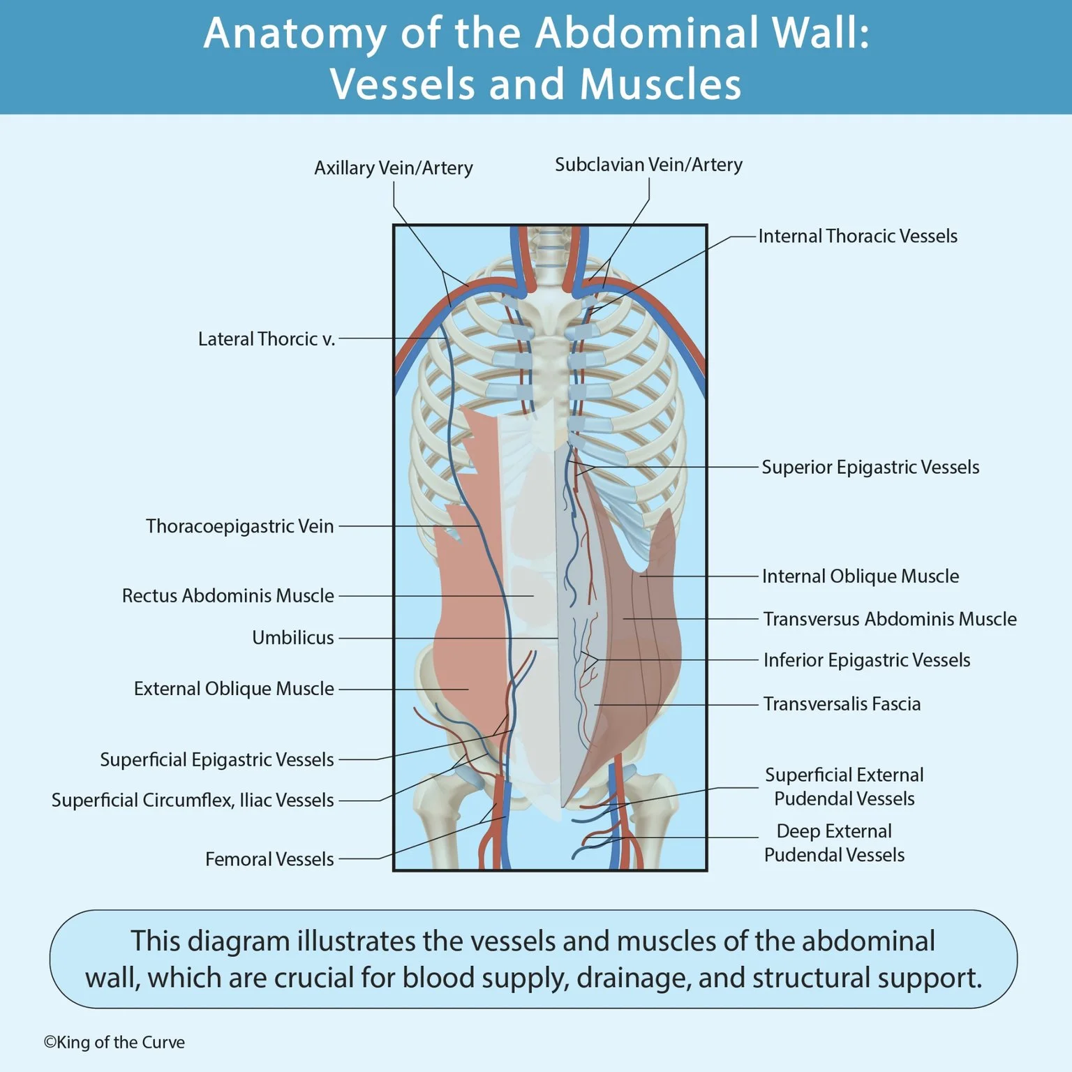
The lateral abdominal wall contains three overlapping muscle layers: the external oblique, internal oblique, and transversus abdominis. From superficial to deep, their fibers generally run downward and medially, upward and medially, and horizontally. These different orientations strengthen the wall while allowing coordinated bending, rotation, and compression.
🧱 Rectus Abdominis and the Rectus Sheath
The paired rectus abdominis muscles run vertically on either side of the midline and help flex the trunk. They lie within the rectus sheath, formed by the broad tendons, or aponeuroses, of the flat abdominal muscles. These aponeuroses meet centrally at the linea alba, which separates the right and left rectus muscles.
🛡️ Where the Transversalis Fascia Fits
The transversalis fascia lies deep to the transversus abdominis muscle and forms part of the internal lining of the abdominal wall. It is distinct from the muscle itself and from the parietal peritoneum farther inward. Below the arcuate line, the rectus abdominis lacks a posterior aponeurotic sheath and rests against the transversalis fascia.
📊 Main Abdominal Wall Muscles and Fascia
Each muscular layer contributes to support and movement, while the fascial layers organize and reinforce the wall. Distinguishing the transversus abdominis from the transversalis fascia is particularly useful because their similar names can cause confusion. The table summarizes their positions and principal roles.
| Structure | Position | Main Role |
|---|---|---|
| External oblique | Most superficial flat muscle | Compresses abdominal contents and assists trunk flexion and opposite-side rotation |
| Internal oblique | Between external oblique and transversus abdominis | Compresses abdominal contents and assists trunk flexion and same-side rotation |
| Transversus abdominis | Deepest flat muscle | Compresses and supports abdominal contents |
| Rectus abdominis | Paired vertical muscles near the midline | Flexes the trunk and helps stabilize the pelvis |
| Rectus sheath | Aponeurotic covering around rectus abdominis | Supports the muscle and contains associated vessels and nerves |
| Transversalis fascia | Deep to transversus abdominis | Forms a supporting fascial layer inside the abdominal wall |
🩸 Superior and Inferior Epigastric Arteries
The superior epigastric artery continues from the internal thoracic artery, while the inferior epigastric artery arises from the external iliac artery. They approach the rectus abdominis from above and below and connect within the rectus sheath. This anastomosis links arterial pathways from the upper and lower regions of the trunk.
🌿 Superficial Blood Supply of the Lower Abdominal Wall
The superficial epigastric and superficial circumflex iliac arteries usually arise from the femoral artery. They supply superficial tissues of the lower abdominal wall and the region near the groin. These vessels differ from the deeper inferior epigastric and deep circumflex iliac arteries, which arise from the external iliac artery.
🔄 Venous Drainage and the Thoracoepigastric Vein
Abdominal wall veins carry blood toward drainage pathways above and below the abdomen. The thoracoepigastric vein connects the superficial epigastric and lateral thoracic venous systems along the side of the trunk. This connection provides a potential collateral route between venous territories associated with the inferior and superior venae cavae.
📍 Why These Anatomical Relationships Matter
The arrangement of muscles, fascia, and vessels helps explain where abdominal wall weaknesses and bleeding can occur. The inferior epigastric vessels also serve as landmarks for distinguishing direct from indirect inguinal hernias. Understanding these relationships supports interpretation of anatomy diagrams, imaging, and the tissue layers encountered during abdominal procedures.

Frequently Asked Questions (FAQs)
-
Aim for 4-6 focused hours, ensuring you incorporate breaks to avoid burnout.
-
Practice mindfulness techniques, take practice exams under realistic conditions, and maintain a balanced lifestyle.
-
Set short-term goals, seek support from mentors, and reward yourself for small achievements.
-
Regular exercise improves focus, reduces stress, and enhances overall mental clarity.
-
KOTC offers personalized learning tools, gamification features, and adaptive question banks to help students stay on track without burnout.