
Sensory receptors detect changes inside or outside the body, including touch, temperature, tissue damage, and body position. They convert a stimulus into an electrical response through a process called sensory transduction. This information travels along sensory pathways toward the central nervous system, where it contributes to perception and coordinated responses.
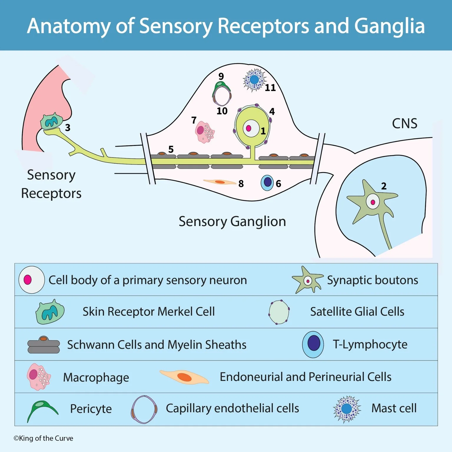
✋ Merkel Cells and Touch Sensation
Merkel cells are specialized cells in the epidermis that work with sensory nerve endings to detect fine details of touch. The Merkel cell–neurite complex responds particularly well to sustained pressure and features such as edges and texture. Its slowly adapting response allows it to continue signaling while a stimulus remains present.
⚡ How Primary Sensory Neurons Carry Signals
Primary sensory neurons carry information from peripheral tissues toward the spinal cord or brainstem. Most neurons in dorsal root ganglia are pseudounipolar, meaning one process leaves the cell body and divides into peripheral and central branches. Signals travel along these branches, while the cell body supports the neuron’s metabolic and maintenance needs.
📍 What Is a Sensory Ganglion?
A sensory ganglion is a collection of sensory neuron cell bodies outside the central nervous system. Dorsal root ganglia contain the cell bodies of spinal sensory neurons, while certain cranial nerve ganglia serve similar functions for the head. Unlike autonomic ganglia, dorsal root ganglia normally do not contain conventional synaptic relays between neurons; information generally continues along the same primary sensory neuron toward the CNS.
📊 Main Cells and Structures in the Sensory Pathway
Sensory signaling depends on neurons working alongside glial, vascular, connective tissue, and immune cells. Each contributes a different function, from detecting mechanical stimuli to maintaining the environment around nerve fibers. The table summarizes the principal structures shown without treating every supporting cell as a sensory receptor.
| Cell or Structure | Main Location | Main Function |
|---|---|---|
| Merkel cell | Epidermis, associated with sensory endings | Contributes to sustained touch and fine-detail detection |
| Primary sensory neuron | Cell body in a sensory ganglion | Carries sensory information toward the CNS |
| Satellite glial cell | Around neuronal cell bodies in peripheral ganglia | Supports neurons and regulates their local environment |
| Schwann cell | Along peripheral axons | Supports nerve fibers and forms myelin around myelinated axons |
| Macrophage | Ganglionic and peripheral nerve tissue | Participates in immune surveillance and debris removal |
| Capillary endothelial cell | Inner lining of small blood vessels | Forms the vascular lining and regulates exchange |
| Pericyte | Along the outer surface of small vessels | Supports vascular stability and function |
🛡️ How Satellite Glial Cells Support Neurons
Satellite glial cells form a close covering around sensory neuron cell bodies. They help regulate the surrounding chemical environment and participate in communication that can influence neuronal excitability. Their position distinguishes them from Schwann cells, which primarily associate with axons rather than forming the sheath around neuronal cell bodies.
🧵 Schwann Cells and Myelin Sheaths
Schwann cells support axons in the peripheral nervous system. Myelinating Schwann cells wrap individual axon segments in insulating layers that allow impulses to travel rapidly between nodes of Ranvier. Other Schwann cells support unmyelinated fibers, so a sensory pathway does not need a thick myelin sheath to transmit information.
🩸 Vascular, Connective Tissue, and Immune Support
Small blood vessels supply sensory ganglia with oxygen and nutrients, while endothelial cells and pericytes contribute to vascular organization. Connective tissue supports the surrounding structures, and immune cells such as macrophages participate in surveillance and responses to injury. Together, these cells create a support system around the sensory neurons.
🔗 Where Sensory Signals Form Synapses
The central branches of primary sensory neurons enter the CNS and connect with other neurons along pathways appropriate to the sensory modality. Some synapse in the spinal cord, while others ascend to the brainstem before their first central relay. A synaptic bouton is an axon terminal specialized for communication; the large, star-shaped structure shown inside the CNS represents a neuron rather than a bouton itself.

Frequently Asked Questions (FAQs)
-
Aim for 4-6 focused hours, ensuring you incorporate breaks to avoid burnout.
-
Practice mindfulness techniques, take practice exams under realistic conditions, and maintain a balanced lifestyle.
-
Set short-term goals, seek support from mentors, and reward yourself for small achievements.
-
Regular exercise improves focus, reduces stress, and enhances overall mental clarity.
-
KOTC offers personalized learning tools, gamification features, and adaptive question banks to help students stay on track without burnout.