
Warfarin is an oral anticoagulant that reduces the blood's ability to form clots by interfering with the vitamin K recycling pathway. It inhibits an enzyme needed to regenerate active vitamin K, preventing the proper activation of several important clotting factors. Understanding warfarin's mechanism connects enzyme inhibition, post-translational modification, blood coagulation, and drug metabolism—important concepts for MCAT biology and biochemistry.
🧬 What Is Warfarin and How Does It Work?
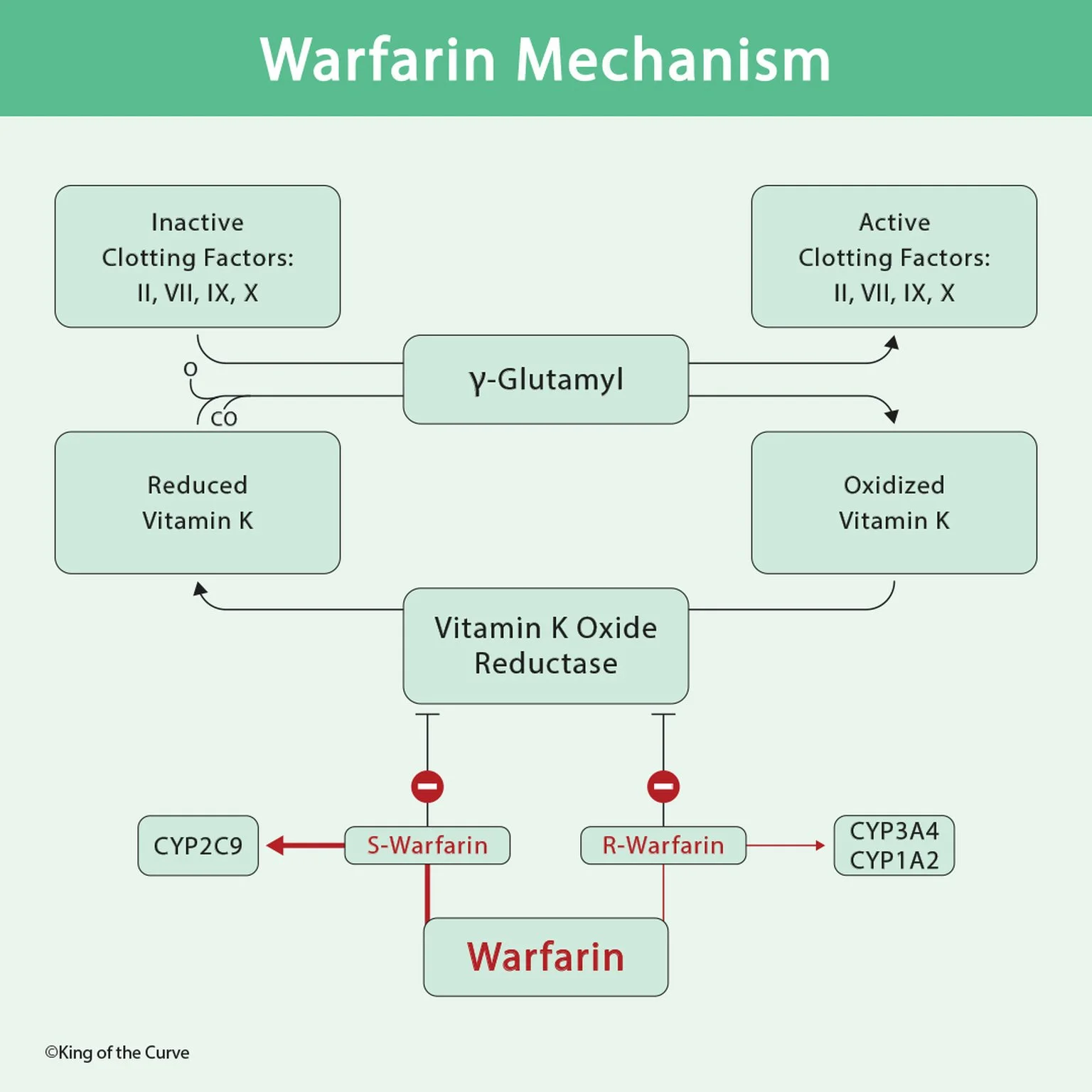
Warfarin is a vitamin K antagonist that reduces the functional activity of vitamin K-dependent coagulation proteins. Its primary target is vitamin K epoxide reductase complex subunit 1 (VKORC1), an enzyme involved in recycling vitamin K.
By inhibiting VKORC1, warfarin decreases the availability of reduced vitamin K, which is required to activate certain clotting proteins. As a result, newly produced vitamin K-dependent clotting factors have impaired coagulation activity. Warfarin does not directly dissolve existing blood clots or immediately inactivate clotting factors already circulating in the bloodstream.
🔄 Understanding the Vitamin K Recycling Pathway
Vitamin K participates in a biochemical cycle that allows it to be reused during clotting factor activation. The reduced form, vitamin K hydroquinone, acts as a cofactor for γ-glutamyl carboxylase. During the carboxylation reaction, vitamin K hydroquinone is oxidized to vitamin K epoxide.
VKORC1 helps recycle vitamin K epoxide into vitamin K quinone, which can subsequently be reduced to vitamin K hydroquinone. Warfarin interrupts this recycling process, limiting the availability of active vitamin K.
⚙️ Gamma-Carboxylation and Clotting Factor Activation
The enzyme γ-glutamyl carboxylase modifies specific glutamate residues in vitamin K-dependent proteins, converting them into γ-carboxyglutamate (Gla) residues. This reaction requires reduced vitamin K, carbon dioxide, and oxygen.
These Gla residues allow clotting factors to bind calcium ions and interact with negatively charged phospholipid membranes, which are essential for efficient coagulation reactions. When warfarin reduces active vitamin K availability, newly synthesized clotting proteins are inadequately carboxylated and have reduced functional activity.
MCAT Tip: Warfarin does not directly inhibit γ-glutamyl carboxylase. It indirectly limits carboxylation by reducing the availability of its vitamin K cofactor.
🩸 Which Clotting Factors Does Warfarin Affect?
Warfarin reduces the functional activity of factors II, VII, IX, and X, all of which require vitamin K-dependent γ-carboxylation. Factor II is prothrombin, factor VII participates in the tissue factor pathway, factor IX functions in the intrinsic pathway, and factor X is part of the common pathway.
A useful mnemonic is 1972, representing factors X, IX, VII, and II. Warfarin also reduces the activity of anticoagulant proteins C and S. Because protein C has a relatively short half-life, warfarin initiation can temporarily create a prothrombotic state before its full anticoagulant effect develops.
💊 S-Warfarin vs. R-Warfarin: Understanding Drug Metabolism
Warfarin is administered as a mixture of two stereoisomers: S-warfarin and R-warfarin. S-warfarin is approximately three to five times more potent as an anticoagulant than R-warfarin and is primarily metabolized by the liver enzyme CYP2C9.
R-warfarin is metabolized through several pathways, including CYP1A2 and CYP3A4. These enzymes belong to the cytochrome P450 family, which participates in the metabolism of many medications. Differences in enzyme activity, genetic variation, and drug interactions can influence warfarin exposure and anticoagulant response.
This makes warfarin a useful example of stereochemistry, enzyme specificity, and pharmacokinetics.
📊 Warfarin Mechanism: High-Yield MCAT Comparison Table
| Component | Primary Function | Relationship to Warfarin |
|---|---|---|
| VKORC1 | Recycles oxidized vitamin K | Directly inhibited |
| Reduced Vitamin K | Cofactor for γ-carboxylation | Availability decreases |
| γ-Glutamyl Carboxylase | Activates vitamin K-dependent proteins | Indirectly limited |
| Factors II, VII, IX, X | Support blood coagulation | Functional activity decreases |
| Proteins C and S | Regulate coagulation | Functional activity decreases |
| S-Warfarin | More potent anticoagulant enantiomer | Primarily metabolized by CYP2C9 |
| R-Warfarin | Less potent anticoagulant enantiomer | Metabolized by CYP1A2, CYP3A4, and other enzymes |
| PT/INR | Measures coagulation pathway activity | Used to monitor warfarin therapy |
Note: VKORC1 is warfarin's pharmacological target, while CYP450 enzymes are responsible for warfarin metabolism. Warfarin indirectly reduces γ-glutamyl carboxylase activity by limiting reduced vitamin K availability.
⏱️ Why Does Warfarin Take Several Days to Work?
Warfarin does not immediately eliminate clotting factors already present in circulation. Instead, its anticoagulant effect develops as existing functional proteins are naturally cleared and replaced by inadequately carboxylated proteins.
Factor VII has a relatively short half-life, allowing early changes in prothrombin time. Factor II has a longer half-life, contributing to the delay in the full antithrombotic effect. This explains why laboratory changes can appear before maximal anticoagulation is achieved.
The delay demonstrates an important pharmacological principle: the biological response to enzyme inhibition may depend on the turnover rate of downstream proteins.
🧪 Warfarin Monitoring: PT, INR, and Drug Interactions
Warfarin therapy is monitored using prothrombin time (PT), reported as the international normalized ratio (INR). PT evaluates the extrinsic and common coagulation pathways and is particularly sensitive to factor VII activity.
Warfarin has a narrow therapeutic index, meaning that relatively small changes in exposure or sensitivity can significantly affect anticoagulation. CYP2C9 inhibitors may increase S-warfarin exposure, while enzyme induction may reduce exposure. Changes in dietary vitamin K intake can also alter the anticoagulant response.
For the MCAT, remember that warfarin is monitored using PT/INR, whereas unfractionated heparin is commonly monitored using aPTT.
🎯 MCAT High-Yield Summary: Remember Warfarin's Mechanism
Warfarin illustrates how enzyme inhibition can disrupt a biochemical recycling pathway and alter protein function. Its central mechanism is:
Warfarin → VKORC1 inhibition → Reduced vitamin K hydroquinone availability → Decreased γ-carboxylation → Reduced functional clotting factors → Anticoagulation.

Frequently Asked Questions (FAQs)
-
Aim for 4-6 focused hours, ensuring you incorporate breaks to avoid burnout.
-
Practice mindfulness techniques, take practice exams under realistic conditions, and maintain a balanced lifestyle.
-
Set short-term goals, seek support from mentors, and reward yourself for small achievements.
-
Regular exercise improves focus, reduces stress, and enhances overall mental clarity.
-
KOTC offers personalized learning tools, gamification features, and adaptive question banks to help students stay on track without burnout.