
Warfarin is an oral anticoagulant that prevents blood clot formation by interfering with the vitamin K cycle, a biochemical pathway essential for producing functional clotting factors. Understanding warfarin's mechanism of action connects biochemistry, enzyme inhibition, blood coagulation, and pharmacology.
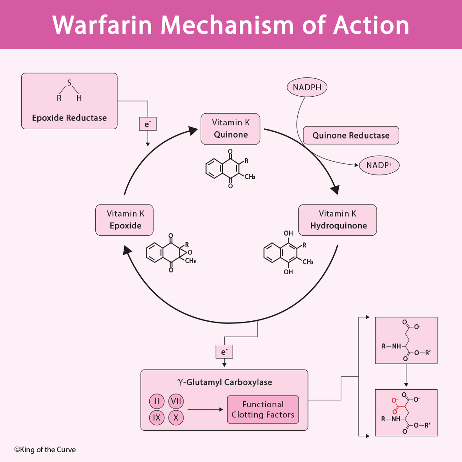
🧬 What Is Warfarin and How Does It Work?
Warfarin is a vitamin K antagonist that reduces the production of functional vitamin K-dependent clotting proteins. Its primary molecular target is vitamin K epoxide reductase complex subunit 1 (VKORC1), a key component of the vitamin K recycling pathway.
Warfarin inhibits VKORC1, reducing the regeneration of vitamin K in its active, reduced form. Without sufficient reduced vitamin K, the liver cannot efficiently activate several proteins involved in coagulation.
Warfarin does not directly dissolve existing blood clots or immediately inactivate clotting factors already circulating in the blood. Instead, it reduces the production of newly synthesized, fully functional vitamin K-dependent proteins.
MCAT Tip: Warfarin is an anticoagulant, not a thrombolytic drug. It helps prevent clot formation and extension rather than directly breaking down existing clots.
🔄 Understanding the Vitamin K Cycle
The vitamin K cycle allows vitamin K to be repeatedly used during the activation of clotting proteins.
Vitamin K exists in several chemical forms, including vitamin K quinone, vitamin K hydroquinone, and vitamin K epoxide.
Vitamin K hydroquinone is the reduced, biologically active cofactor required for γ-glutamyl carboxylase activity.
During carboxylation, vitamin K hydroquinone is oxidized to vitamin K epoxide. The vitamin K recycling system then converts the epoxide back toward the reduced, active form.
🧪 Vitamin K Epoxide Reductase: Warfarin's Primary Target
Vitamin K epoxide reductase is essential for regenerating usable vitamin K from its oxidized epoxide form.
The enzyme complex, whose catalytic component is encoded by VKORC1, reduces vitamin K epoxide to vitamin K quinone and can also participate in further reduction toward vitamin K hydroquinone.
Warfarin inhibits this pathway, limiting the amount of active vitamin K available for γ-carboxylation.
Other vitamin K reductase pathways can contribute to vitamin K reduction, which helps explain why vitamin K administration can counteract warfarin's anticoagulant effects.
This is an important biochemical example of enzyme inhibition altering the availability of a required cofactor.
⚙️ Gamma-Carboxylation: Why Vitamin K Is Essential for Clotting
Vitamin K hydroquinone serves as a cofactor for γ-glutamyl carboxylase, an enzyme that modifies specific glutamate residues in clotting proteins.
This post-translational modification converts selected glutamate residues into γ-carboxyglutamate (Gla) residues.
The reaction can be summarized as:
This modification enables clotting proteins to bind calcium ions effectively.
Calcium binding allows these proteins to interact with negatively charged phospholipid surfaces, particularly those exposed on activated platelets, supporting efficient coagulation reactions.
Without sufficient γ-carboxylation, the proteins may still be synthesized but have substantially reduced coagulation activity.
High-yield concept: Warfarin decreases clotting factor function by preventing adequate post-translational activation, rather than directly stopping synthesis of the clotting factor proteins.
🩸 Which Clotting Factors Does Warfarin Affect?
Warfarin reduces the functional activity of four vitamin K-dependent coagulation factors:
Factor II: Prothrombin, the precursor of thrombin.
Factor VII: Participates in the tissue factor-dependent initiation of coagulation.
Factor IX: Functions in the intrinsic pathway.
Factor X: Participates in the common coagulation pathway.
An easy way to remember these factors is 1972, representing factors X, IX, VII, and II when read as 10, 9, 7, and 2.
Warfarin also affects the vitamin K-dependent anticoagulant proteins C and S, which normally help limit excessive coagulation.
This creates an important clinical consideration: protein C activity can decrease relatively quickly after warfarin initiation, sometimes producing a temporary prothrombotic state before the full anticoagulant effect develops.
📊 Warfarin Mechanism of Action: High-Yield MCAT Table
The following table summarizes the major biochemical components involved in warfarin's anticoagulant mechanism.
| Component | Normal Function | Effect of Warfarin |
|---|---|---|
| VKORC1 | Recycles vitamin K epoxide toward reduced vitamin K | Inhibited |
| Vitamin K Hydroquinone | Cofactor for γ-carboxylation | Reduced availability |
| γ-Glutamyl Carboxylase | Carboxylates glutamate residues | Activity limited by reduced cofactor availability |
| Factors II, VII, IX, X | Support blood coagulation | Reduced functional activity |
| Proteins C and S | Regulate and inhibit coagulation | Reduced functional activity |
| Calcium Binding | Enables clotting proteins to associate with phospholipid surfaces | Impaired in undercarboxylated proteins |
| PT/INR | Assesses clotting through the extrinsic and common pathways | Typically increases with anticoagulation |
Note: Warfarin does not directly inhibit γ-glutamyl carboxylase or calcium binding. These effects occur indirectly because reduced vitamin K becomes less available. PT/INR is a monitoring measurement, not a molecular target of warfarin.
⏱️ Why Does Warfarin Have a Delayed Anticoagulant Effect?
Warfarin does not immediately eliminate functional clotting factors already present in circulation.
Its anticoagulant effect develops as previously synthesized clotting factors are naturally cleared and replaced by proteins with inadequate γ-carboxylation.
Different clotting factors have different circulating half-lives. Factor VII has a relatively short half-life, so changes in the prothrombin time may occur before the full anticoagulant effect is established.
Factor II (prothrombin) has a longer half-life, contributing to the delay in achieving the full antithrombotic effect.
For this reason, warfarin's anticoagulant action develops over several days, even though laboratory changes may appear earlier.
This distinction illustrates how protein turnover and enzyme inhibition can influence the timing of a drug's physiological effects.
🧫 Warfarin Monitoring: PT, INR, and Vitamin K
Warfarin therapy is monitored primarily using the prothrombin time (PT), standardized as the international normalized ratio (INR).
PT evaluates the extrinsic and common coagulation pathways and is particularly sensitive to changes in factor VII activity.
The INR helps standardize PT measurements between laboratories.
A higher INR generally indicates a longer clotting time and a greater degree of anticoagulation, although bleeding risk depends on additional clinical factors.
Vitamin K can be administered to reverse warfarin's effects, while urgent reversal in serious bleeding may require additional treatment such as four-factor prothrombin complex concentrate.
For the MCAT, the key distinction is that warfarin is monitored using PT/INR, whereas unfractionated heparin is commonly monitored using aPTT.
🎯 MCAT High-Yield Summary: Remember Warfarin's Mechanism
Warfarin is a classic example of how disrupting a biochemical recycling pathway can change the function of an entire physiological system.
Remember the sequence:
Warfarin → VKORC1 inhibition → Reduced active vitamin K → Decreased γ-carboxylation → Reduced functional clotting factors → Anticoagulation.

Frequently Asked Questions (FAQs)
-
Aim for 4-6 focused hours, ensuring you incorporate breaks to avoid burnout.
-
Practice mindfulness techniques, take practice exams under realistic conditions, and maintain a balanced lifestyle.
-
Set short-term goals, seek support from mentors, and reward yourself for small achievements.
-
Regular exercise improves focus, reduces stress, and enhances overall mental clarity.
-
KOTC offers personalized learning tools, gamification features, and adaptive question banks to help students stay on track without burnout.