
🧬 Understanding Vitelline Duct Remnants for the MCAT
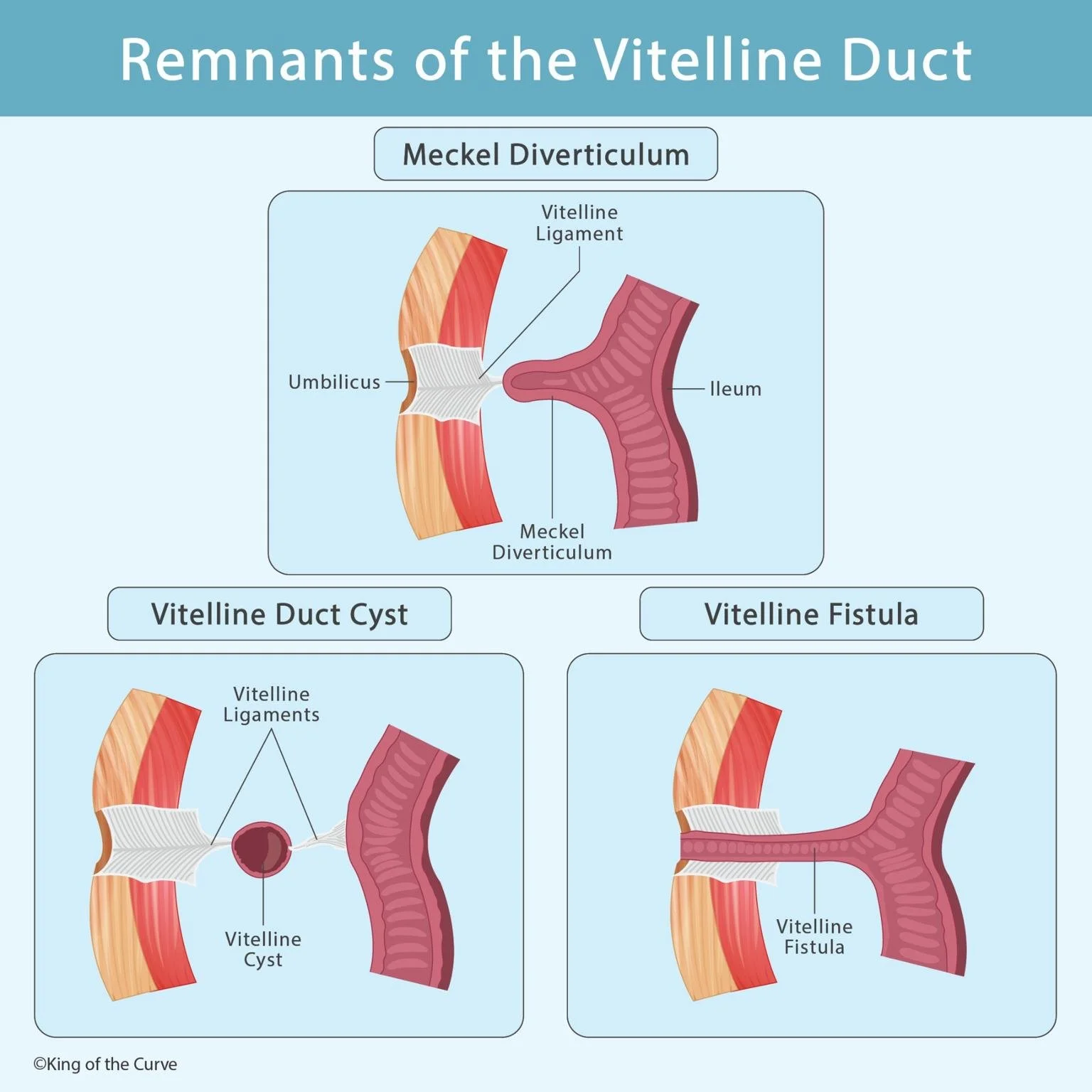
During early embryologic development, the vitelline duct connects the midgut to the yolk sac. Normally, this duct completely obliterates by the 7th week of gestation. But when it doesn’t, several clinically relevant anatomic anomalies can occur many of which appear on MCAT passages and NBME-style questions. King of the Curve’s visual highlights the three major remnants: Meckel diverticulum, vitelline duct cyst, and vitelline fistula.
🩺 Meckel Diverticulum: The Most Common Remnant
Meckel diverticulum is the most common congenital anomaly of the GI tract. It forms when the proximal portion of the vitelline duct persists, creating a true diverticulum of the ileum. A classic high-yield detail: it may contain ectopic gastric or pancreatic tissue, leading to lower GI bleeding in children. The KOTC image shows the diverticulum projecting from the ileum toward the umbilicus, attached by a fibrous vitelline ligament.
🫙 Vitelline Duct Cyst: Persistence of the Middle Portion
A vitelline duct cyst (also called an enterocyst) occurs when the central portion of the duct remains while both ends close. This creates a fibrous band between the ileum and umbilicus, with a fluid-filled cyst suspended in the middle. On the MCAT, this anomaly is relevant because the connecting bands may cause intestinal obstruction or volvulus.
🔗 Vitelline Fistula: A Complete Persistence
A vitelline fistula results when the entire duct remains patent, forming a direct connection between the ileum and the umbilicus. This leads to the hallmark symptom: meconium discharge from the umbilicus in newborns. The KOTC graphic highlights this continuous channel, making it easy to visualize how stool can reach the abdominal wall.
📊 High-Yield Comparison Table: Vitelline Duct Remnants
| Remnant | What Persists? | Key Symptoms | MCAT Clues |
|---|---|---|---|
| Meckel Diverticulum | Proximal duct | Painless GI bleeding; ectopic tissue | “Rule of 2s,” technetium scan |
| Vitelline Duct Cyst | Middle duct only | Possible intestinal obstruction | Cyst attached by fibrous ligaments |
| Vitelline Fistula | Entire duct | Stool leakage from umbilicus | Persistent connection to ileum |
🧠 Why the MCAT Loves This Topic
Vitelline duct remnants sit at the intersection of embryology, anatomy, and clinical presentation—making them perfect for MCAT integration. Many questions require identifying the anomaly from symptoms alone or predicting complications like bleeding, infection, or obstruction. Understanding the embryologic origin helps you reason through even unfamiliar presentations.
📚 Mastering Embryology With KOTC Visuals
Embryology can feel abstract, but KOTC visuals transform it into digestible, memorable images. Pairing diagrams like this one with the Adaptive Q-Bank, Daily Questions, and structured study tools at kingofthecurve.org/studyscience helps reinforce long-term memory and boosts your confidence on test day. When you visualize development clearly, the MCAT becomes far more predictable.

Frequently Asked Questions (FAQs)
-
Aim for 4-6 focused hours, ensuring you incorporate breaks to avoid burnout.
-
Practice mindfulness techniques, take practice exams under realistic conditions, and maintain a balanced lifestyle.
-
Set short-term goals, seek support from mentors, and reward yourself for small achievements.
-
Regular exercise improves focus, reduces stress, and enhances overall mental clarity.
-
KOTC offers personalized learning tools, gamification features, and adaptive question banks to help students stay on track without burnout.