🧠 Spinal Cord Anatomy: Structure, Regions & MCAT Essentials
🧬 What Is the Spinal Cord?
The spinal cord is a major component of the central nervous system (CNS). It extends inferiorly from the medulla oblongata through the vertebral canal and provides a pathway for communication between the brain and the body. It also plays a central role in reflexes.
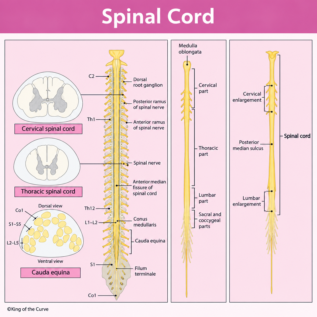
📍 Major Regions of the Spinal Cord
The spinal cord is divided into cervical, thoracic, lumbar, sacral, and coccygeal regions. Together, these regions give rise to 31 pairs of spinal nerves: 8 cervical, 12 thoracic, 5 lumbar, 5 sacral, and 1 coccygeal pair.
🔬 Gray Matter and White Matter
In cross-section, the spinal cord contains butterfly-shaped gray matter surrounded by white matter. Gray matter contains many neuronal cell bodies and synapses, whereas white matter contains ascending and descending axonal tracts that transmit information between the spinal cord and brain.
🔄 Dorsal Roots, Ventral Roots, and Spinal Nerves
The dorsal root carries sensory—or afferent—information toward the spinal cord, while the ventral root carries motor—or efferent—information away from it. The dorsal root ganglion contains the cell bodies of primary sensory neurons. The dorsal and ventral roots unite to form a mixed spinal nerve.
💪 Cervical and Lumbosacral Enlargements
The cervical enlargement contains additional neural tissue associated with the upper limbs, while the lumbosacral enlargement is associated with the lower limbs. These enlargements reflect the greater sensory and motor innervation required by the extremities.
🐴 Conus Medullaris and Cauda Equina
The adult spinal cord usually terminates around the L1–L2 vertebral level, forming the tapered conus medullaris. Nerve roots continuing downward below this point form the cauda equina. The filum terminale is a thin fibrous extension that helps anchor the spinal cord.
| Structure | Description | High-Yield Function |
|---|---|---|
| Dorsal Root | Posterior spinal nerve root | Carries sensory signals toward the spinal cord |
| Dorsal Root Ganglion | Collection of sensory neuron cell bodies | Contains primary sensory neuron cell bodies |
| Ventral Root | Anterior spinal nerve root | Carries motor signals away from the spinal cord |
| Spinal Nerve | Union of dorsal and ventral roots | Contains both sensory and motor fibers |
| Cervical Enlargement | Enlarged cervical region | Associated with upper-limb innervation |
| Lumbosacral Enlargement | Enlarged lower spinal cord | Associated with lower-limb innervation |
| Conus Medullaris | Tapered terminal portion of the spinal cord | Marks the inferior end of the spinal cord |
| Cauda Equina | Bundle of descending spinal nerve roots | Supplies lower-body regions |
| Filum Terminale | Thin fibrous extension | Helps anchor the spinal cord |
⚡ Reflexes and Neural Pathways
The spinal cord can coordinate reflexes without requiring conscious processing before the response occurs. Sensory information enters through the dorsal side, while motor output exits through the ventral side. At the same time, ascending tracts carry sensory signals toward the brain and descending tracts carry motor commands toward the spinal cord.
🎯 MCAT High-Yield Takeaway
Remember the core pattern: dorsal = sensory, ventral = motor. Know the cervical and lumbosacral enlargements, conus medullaris, cauda equina, dorsal root ganglion, and the difference between gray and white matter. These concepts commonly connect with MCAT questions about reflexes, sensory pathways, motor control, and spinal cord lesions.