
Transposition of the Great Vessels (TGV): A High-Yield MCAT Guide
🧬 What Is Transposition of the Great Vessels?
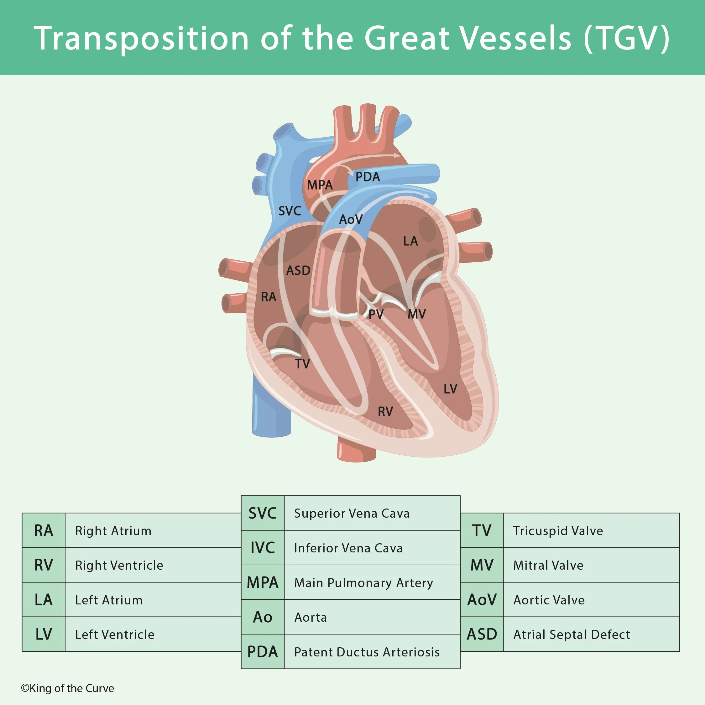
Transposition of the great vessels, more commonly called transposition of the great arteries (TGA), is a congenital heart defect in which the aorta arises from the right ventricle and the pulmonary artery arises from the left ventricle. This creates two parallel circulatory loops instead of the normal series circulation.
🔄 How Does Normal Circulation Differ?
Normally, deoxygenated blood flows from the right ventricle → pulmonary artery → lungs, while oxygenated blood flows from the left ventricle → aorta → systemic circulation. In TGA, these connections are reversed. The right ventricle sends deoxygenated blood back to the body, while the left ventricle repeatedly sends oxygenated blood back to the lungs.
🩸 Why Is Blood Mixing Essential?
Because the pulmonary and systemic circulations run in parallel, survival requires some communication between them. Mixing may occur through an atrial septal defect (ASD), ventricular septal defect (VSD), or patent ductus arteriosus (PDA). These connections allow oxygenated and deoxygenated blood to mix, improving systemic oxygen delivery.
👶 Clinical Presentation
Newborns with significant TGA commonly develop cyanosis shortly after birth. The severity depends largely on how much mixing occurs between the two circulations. When communication is limited, oxygen saturation can remain dangerously low despite supplemental oxygen because the underlying problem is abnormal circulation rather than inadequate ventilation.
💊 Role of the Patent Ductus Arteriosus
A PDA can temporarily improve oxygenation by providing another route for blood mixing. In a critically cyanotic newborn with suspected TGA, prostaglandin E₁ may be used clinically to maintain ductal patency until definitive intervention can be performed. For exam purposes, connect prostaglandin E₁ with keeping the ductus arteriosus open.
| 🫀 Feature | ✅ Normal Heart | 🔄 Transposition of Great Arteries |
|---|---|---|
| RV outflow | Pulmonary artery | Aorta |
| LV outflow | Aorta | Pulmonary artery |
| Circulation pattern | Series | Parallel |
| Oxygenated blood reaches body | Directly | Requires mixing |
| Helpful communication | Not required | ASD, VSD, or PDA |
🧠 Embryology Connection
TGA is classically associated with abnormal development of the aorticopulmonary septum. Normally, this septum spirals during embryogenesis so that the aorta connects with the left ventricle and the pulmonary trunk connects with the right ventricle. Failure of appropriate spiraling can produce the abnormal ventricular–arterial connections seen in TGA.
🩺 Treatment Principles
Initial management focuses on maintaining adequate mixing and oxygen delivery. If atrial-level mixing is inadequate, a procedure such as balloon atrial septostomy may be used to enlarge the atrial communication. Definitive correction commonly involves an arterial switch operation, which reconnects the great arteries to the appropriate ventricles.
🎯 MCAT High-Yield Takeaway
For the MCAT, remember the core relationship: TGA = aorta from RV + pulmonary artery from LV → parallel circulation → severe neonatal cyanosis → survival depends on mixing. Think of ASD, VSD, or PDA as beneficial shunts in this setting, and remember that prostaglandin E₁ keeps the PDA open.