
🧠 Sensory Receptors and Ganglion Structure Explained
The nervous system constantly gathers information from the environment and transmits it to the brain. This process begins at sensory receptors and passes through specialized structures known as sensory ganglia before reaching the central nervous system (CNS).
👋 Sensory Receptors: Where It All Begins
Sensory receptors detect changes in the environment such as:
Touch
Pressure
Temperature
Pain
Vibration
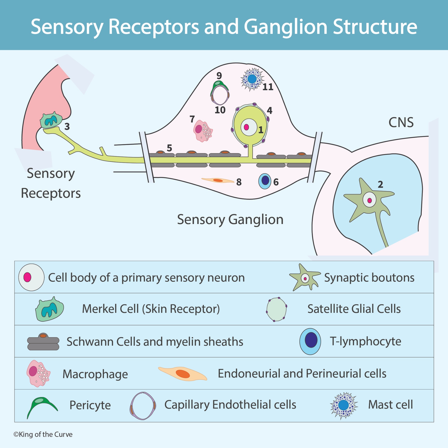
In the image, a Merkel cell (skin receptor) is shown interacting with the terminal of a primary sensory neuron. Merkel cells are responsible for detecting fine touch and pressure.
When stimulated, the receptor generates an electrical signal that travels along the sensory neuron.
🔗 Primary Sensory Neuron Structure
A primary sensory neuron is typically pseudounipolar, meaning it has:
A peripheral process (toward the receptor)
A central process (toward the CNS)
A single cell body located in a sensory ganglion
The cell body of the primary sensory neuron is located in the sensory ganglion — not inside the CNS.
This arrangement allows signals to bypass synapses in the ganglion and travel directly into the spinal cord or brainstem.
🏢 The Sensory Ganglion
A sensory ganglion (such as a dorsal root ganglion) contains:
Neuronal cell bodies
Satellite glial cells
Blood vessels
Immune cells
Connective tissue cells
Unlike autonomic ganglia, sensory ganglia do not contain synapses between neurons.
🧬 Satellite Glial Cells
Each sensory neuron cell body is surrounded by satellite glial cells.
Their functions include:
Structural support
Nutrient exchange
Regulation of the extracellular environment
Protection of neurons
They play a role similar to astrocytes in the CNS.
🧵 Schwann Cells and Myelin
Along the axon, Schwann cells form the myelin sheath.
Myelin allows:
Faster signal conduction
Saltatory conduction
Efficient transmission of sensory information
These cells are essential for peripheral nerve function.
🛡 Immune and Support Cells in the Ganglion
The ganglion also contains:
Macrophages – immune surveillance and debris removal
T-lymphocytes – immune response regulation
Mast cells – inflammatory response
Pericytes – support of blood vessels
Capillary endothelial cells – line blood vessels
Endoneurial and perineurial cells – connective tissue layers that protect nerve fibers
These cells maintain the health and integrity of the peripheral nervous system.
📋 Components of Sensory Receptors and Ganglion Structure
| Component | Location | Function | Clinical Relevance |
|---|---|---|---|
| Merkel Cell (Skin Receptor) | Skin (epidermis) | Detects fine touch and pressure | Important in tactile sensation disorders |
| Primary Sensory Neuron Cell Body | Sensory ganglion (e.g., dorsal root ganglion) | Houses nucleus of pseudounipolar neuron | Affected in herpes zoster (shingles) |
| Peripheral Process | Extends from receptor to ganglion | Carries sensory signals toward cell body | Damage leads to sensory loss |
| Central Process | Extends from ganglion to CNS | Transmits signals to spinal cord/brainstem | Involved in sensory pathways |
| Satellite Glial Cells | Surround neuron cell bodies in ganglion | Structural support and metabolic regulation | Implicated in neuropathic pain |
| Schwann Cells | Along peripheral axons | Form myelin sheath for rapid conduction | Damaged in peripheral neuropathies |
| Macrophages | Within ganglion | Immune surveillance and debris removal | Active during nerve injury |
| T-Lymphocytes | Within ganglion | Immune response regulation | Involved in inflammatory neuropathies |
| Mast Cells | Near blood vessels | Mediate inflammation | Contribute to neuroinflammatory pain |
| Pericytes | Around capillaries | Support blood vessel stability | Important in vascular regulation |
| Capillary Endothelial Cells | Blood vessel lining | Form blood-nerve barrier | Barrier dysfunction can cause edema |
| Endoneurial & Perineurial Cells | Connective tissue layers of nerve | Protect and structurally organize nerve fibers | Damage affects nerve integrity |
🚪 Entry into the CNS
The central process of the primary sensory neuron enters the CNS, where it synapses with second-order neurons.
This is where:
Signal integration occurs
Reflex pathways begin
Sensory information ascends to higher centers
🧠 Why This Structure Matters
Understanding sensory ganglion structure is important for:
Neuropathic pain disorders
Peripheral neuropathies
Herpes zoster (shingles)
Inflammatory nerve conditions
Nerve injury recovery
Damage to any of these components can alter sensation or cause chronic pain.
📌 Key Takeaways
Sensory neurons are pseudounipolar.
Their cell bodies reside in sensory ganglia.
Sensory ganglia do not contain synapses.
Satellite glial cells surround neuronal cell bodies.
Schwann cells myelinate peripheral axons.
Immune and vascular cells support ganglion health.

Frequently Asked Questions (FAQs)
-
Aim for 4-6 focused hours, ensuring you incorporate breaks to avoid burnout.
-
Practice mindfulness techniques, take practice exams under realistic conditions, and maintain a balanced lifestyle.
-
Set short-term goals, seek support from mentors, and reward yourself for small achievements.
-
Regular exercise improves focus, reduces stress, and enhances overall mental clarity.
-
KOTC offers personalized learning tools, gamification features, and adaptive question banks to help students stay on track without burnout.