
Ischemic colitis develops when blood flow to part of the colon becomes insufficient to meet its needs. The resulting shortage of oxygen can injure the intestinal lining, causing inflammation, abdominal pain, and bleeding. Many cases involve temporary injury, but severe ischemia can damage the full thickness of the bowel wall.
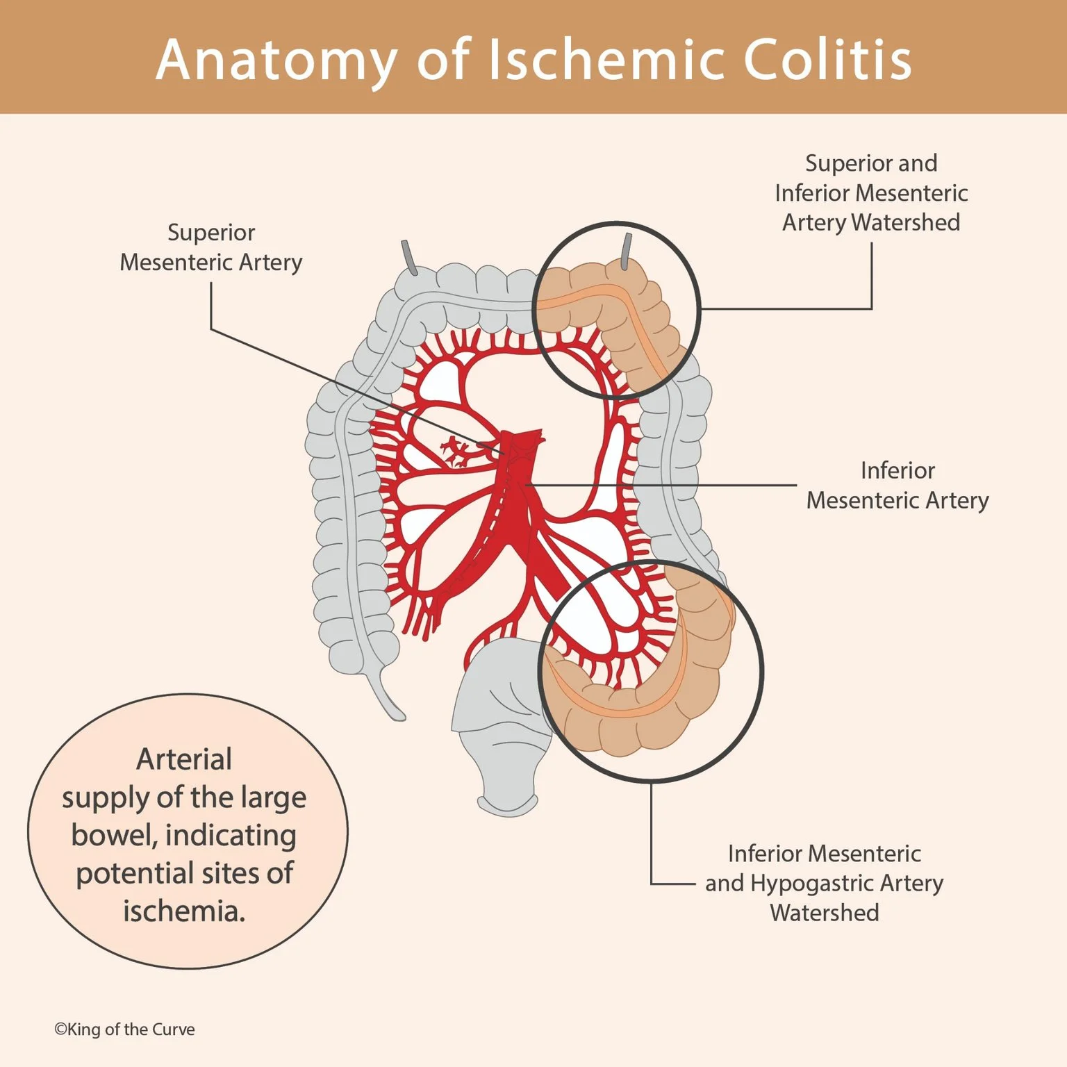
🌳 How the Colon Receives Its Blood Supply
The superior mesenteric artery supplies the cecum, ascending colon, and approximately the proximal two-thirds of the transverse colon. The inferior mesenteric artery supplies the distal transverse colon, descending colon, sigmoid colon, and upper rectum through its branches. Connections between these vessels provide collateral routes that help maintain blood flow.
📍 What Makes a Watershed Area Vulnerable?
A watershed area lies near the boundary between arterial territories, where collateral circulation may be less robust. These regions can become especially vulnerable when blood pressure or circulating volume falls. The splenic flexure and rectosigmoid junction are classic examples, although ischemic injury can affect other parts of the colon.
↪️ The Splenic Flexure and Griffith’s Point
The splenic flexure is the bend between the transverse and descending colon near the spleen. Its blood supply lies at the junction of territories supplied by the middle colic branch of the superior mesenteric artery and the left colic branch of the inferior mesenteric artery. This watershed region, commonly called Griffith’s point, is vulnerable when connecting vessels cannot provide adequate flow.
📊 Key Arterial Territories and Watershed Regions
The location of ischemic injury becomes easier to understand when each bowel segment is connected with its arterial supply. The marginal artery provides an important connection along the colon, but its development varies between individuals. The table highlights the major territories and the two classic watershed regions.
| Region | Main Arterial Supply | Anatomical Significance |
|---|---|---|
| Cecum and ascending colon | Superior mesenteric artery branches | Right-sided territory that can also be vulnerable during low-flow states |
| Proximal two-thirds of transverse colon | Middle colic artery, from the superior mesenteric artery | Connects with left colic circulation near the splenic flexure |
| Splenic flexure | Middle colic and left colic territories | Classic watershed region at Griffith’s point |
| Descending and sigmoid colon | Left colic and sigmoid branches of the inferior mesenteric artery | Major left-colon arterial territory |
| Rectosigmoid junction | Last sigmoid and superior rectal arterial territories | Classic watershed region at Sudeck’s point |
| Rectum | Superior rectal artery plus internal iliac–derived contributions | Additional collateral supply helps explain relative rectal sparing |
🔗 The Rectosigmoid Junction and Sudeck’s Point
Sudeck’s point refers to the potentially vulnerable connection between the last sigmoid artery and the superior rectal artery, both arising from the inferior mesenteric system. It should not be defined simply as an inferior mesenteric–internal iliac boundary. Internal iliac–derived rectal vessels provide additional collateral circulation farther along the rectum, helping protect it from ischemia.
📉 Why Blood Flow Can Become Insufficient
Ischemic colitis often occurs without a complete blockage of a major artery. Reduced circulating volume, low blood pressure, impaired cardiac output, or narrowing of smaller vessels can compromise colonic perfusion. Older age, vascular disease, certain medications, and procedures involving the aorta may increase susceptibility, depending on the clinical situation.
🚨 Recognizing Symptoms and Serious Complications
A typical presentation includes sudden cramping abdominal pain, an urgent need to pass stool, and subsequent rectal bleeding or bloody diarrhea. However, symptoms vary, and right-sided disease may cause significant pain without obvious bleeding. Severe or worsening abdominal pain, fainting, or substantial bleeding requires urgent medical assessment because bowel injury can progress.
🩺 How Ischemic Colitis Is Evaluated and Managed
Clinicians combine the history and examination with investigations such as blood tests, contrast-enhanced CT, and, when appropriate, lower gastrointestinal endoscopy. Treatment depends on severity and may include intravenous fluids, bowel rest, correction of the underlying cause, and antibiotics in selected cases. Surgery may be necessary when there is bowel gangrene, perforation, or other evidence of severe injury.

Frequently Asked Questions (FAQs)
-
Aim for 4-6 focused hours, ensuring you incorporate breaks to avoid burnout.
-
Practice mindfulness techniques, take practice exams under realistic conditions, and maintain a balanced lifestyle.
-
Set short-term goals, seek support from mentors, and reward yourself for small achievements.
-
Regular exercise improves focus, reduces stress, and enhances overall mental clarity.
-
KOTC offers personalized learning tools, gamification features, and adaptive question banks to help students stay on track without burnout.