
🩺 Nephrotic Syndrome Summary: Causes, Pathology, Diagnosis, and Treatment
Nephrotic syndrome is a clinical condition characterized by massive proteinuria, hypoalbuminemia, generalized edema, hyperlipidemia, and lipiduria. It results from increased permeability of the glomerular filtration barrier, allowing proteins primarily albumin to leak into the urine.
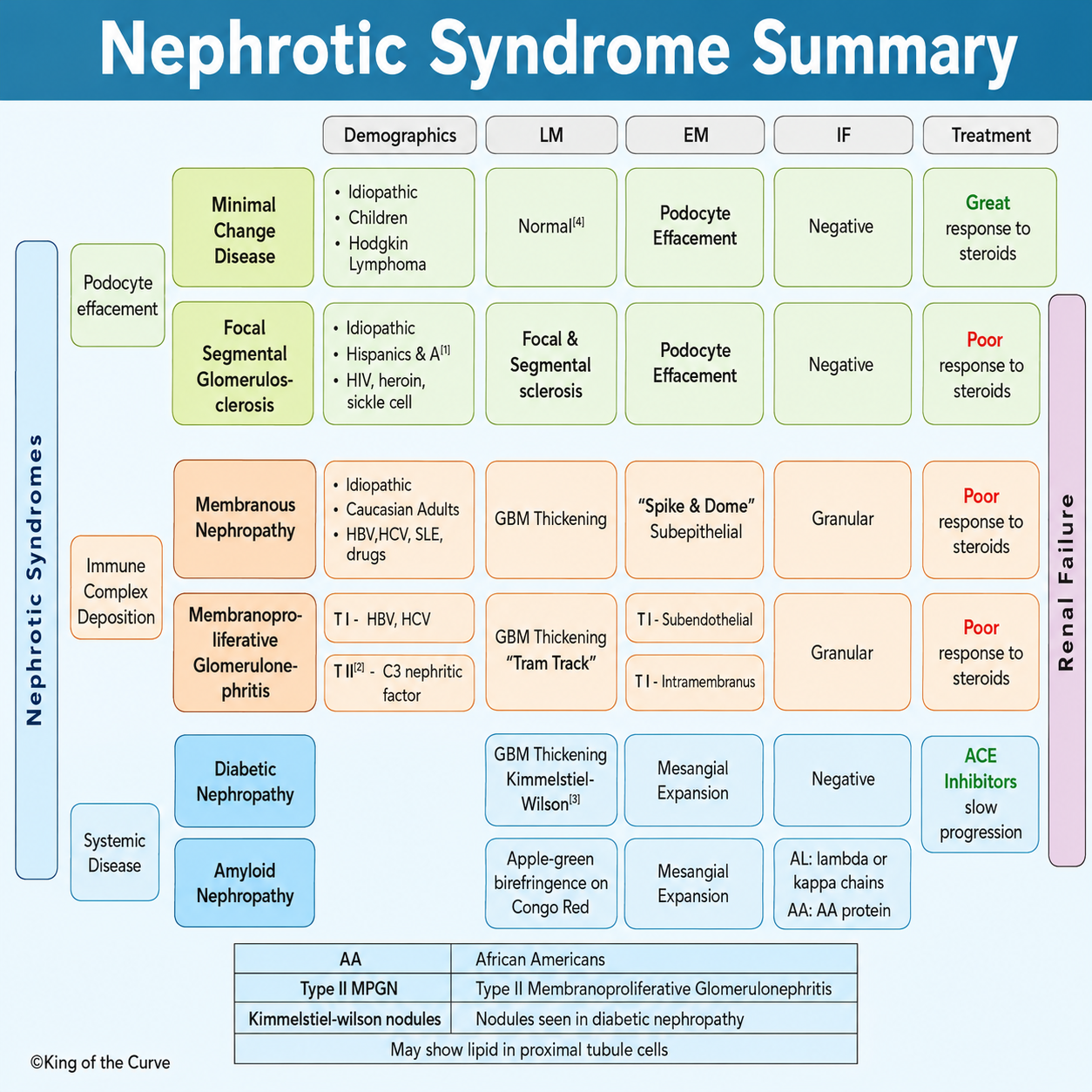
📌 What Is Nephrotic Syndrome?
Nephrotic syndrome occurs when injury to the glomerular capillary wall causes excessive protein loss into the urine.
Hallmark Clinical Features
Proteinuria (>3.5 g/day)
Hypoalbuminemia
Generalized edema
Hyperlipidemia
Lipiduria (fatty casts)
Patients are also at increased risk of thrombosis and infections due to the urinary loss of anticoagulant and immune proteins.
🟢 Podocyte Effacement Disorders
These disorders primarily affect the podocytes, leading to widespread foot process effacement.
🔹 Minimal Change Disease (MCD)
Minimal Change Disease is the most common cause of nephrotic syndrome in children.
Common Associations
Idiopathic
Children
Hodgkin lymphoma
Microscopy Findings
Light Microscopy (LM)
Normal glomeruli
Electron Microscopy (EM)
Diffuse podocyte foot process effacement
Immunofluorescence (IF)
Negative
Treatment
Excellent response to corticosteroids
Prognosis
Most children achieve complete remission with steroid therapy.
🔹 Focal Segmental Glomerulosclerosis (FSGS)
FSGS is a common cause of nephrotic syndrome in adults and frequently progresses to chronic kidney disease.
Common Associations
Idiopathic
HIV infection
Heroin use
Sickle cell disease
Higher prevalence in individuals of African ancestry
Microscopy Findings
LM
Focal and segmental sclerosis
EM
Podocyte foot process effacement
IF
Usually negative
Treatment
Limited response to steroids
May require calcineurin inhibitors or immunosuppressive therapy
Prognosis
Often progresses to chronic kidney disease and renal failure.
🟠 Immune Complex–Mediated Disorders
These conditions result from immune complex deposition within the glomerulus.
🔹 Membranous Nephropathy
Membranous nephropathy is one of the most common causes of nephrotic syndrome in adults.
Common Associations
Idiopathic
Hepatitis B
Hepatitis C
Systemic lupus erythematosus (SLE)
Certain medications
Microscopy Findings
LM
Thickened glomerular basement membrane (GBM)
EM
Subepithelial immune deposits
Classic "spike and dome" appearance
IF
Granular staining
Treatment
Conservative management
Immunosuppressive therapy in selected patients
Prognosis
Variable; some patients progress to chronic kidney disease.
🔹 Membranoproliferative Glomerulonephritis (MPGN)
MPGN is characterized by mesangial proliferation and immune-mediated injury.
Types
Type I
Associated with hepatitis B and hepatitis C
Type II (Dense Deposit Disease)
Associated with C3 nephritic factor
Microscopy Findings
LM
GBM thickening
Tram-track appearance
EM
Type I: Subendothelial deposits
Type II: Intramembranous dense deposits
IF
Granular immune deposits
Treatment
Management focuses on treating the underlying cause and controlling disease progression.
🔵 Systemic Disease–Associated Nephropathies
🔹 Diabetic Nephropathy
The leading cause of chronic kidney disease worldwide.
Characteristic Findings
LM
GBM thickening
Kimmelstiel–Wilson nodules
EM
Mesangial expansion
IF
Negative
Treatment
ACE inhibitors
ARBs
Tight glycemic control
Prognosis
ACE inhibitors and ARBs slow disease progression.
🔹 Amyloid Nephropathy
Amyloid deposition within the kidneys causes nephrotic syndrome and progressive renal dysfunction.
Microscopy Findings
LM
Congo red-positive deposits
Apple-green birefringence under polarized light
EM
Mesangial expansion
IF Findings
Depends on amyloid subtype:
AL amyloidosis → Lambda or kappa light chains
AA amyloidosis → AA protein
Treatment
Treat the underlying cause of amyloid production.
🔬 Light Microscopy, Electron Microscopy, and Immunofluorescence Patterns
| 🩺 Disease | 🔬 Light Microscopy (LM) | 🧫 Electron Microscopy (EM) | ✨ Immunofluorescence (IF) |
|---|---|---|---|
| Minimal Change Disease | Normal | Podocyte foot process effacement | Negative |
| Focal Segmental Glomerulosclerosis (FSGS) | Focal and segmental sclerosis | Podocyte foot process effacement | Usually negative |
| Membranous Nephropathy | GBM thickening | Subepithelial deposits | Granular |
| Membranoproliferative Glomerulonephritis (MPGN) | Tram-track GBM appearance | Subendothelial or intramembranous deposits | Granular |
| Diabetic Nephropathy | Kimmelstiel–Wilson nodules | Mesangial expansion | Negative |
| Amyloidosis | Congo red-positive deposits | Amyloid fibrils | Variable |
💊 Treatment Overview
Treatment depends on the underlying pathology.
Minimal Change Disease: Corticosteroids
FSGS: Steroids or immunosuppressants
Membranous Nephropathy: Supportive care ± immunosuppression
MPGN: Treat underlying infection or complement disorder
Diabetic Nephropathy: ACE inhibitors, ARBs, glycemic control
Amyloidosis: Disease-specific therapy
⚠️ High-Yield USMLE Pearls
Minimal Change Disease → Children + steroid responsive + normal LM
FSGS → HIV, heroin, sickle cell disease, African ancestry
Membranous Nephropathy → Spike-and-dome appearance
MPGN → Tram-track GBM
Diabetic Nephropathy → Kimmelstiel–Wilson nodules
Amyloidosis → Congo red stain with apple-green birefringence
🎯 Key Takeaways
Nephrotic syndrome encompasses several diseases with distinct pathological mechanisms but similar clinical manifestations. Differentiating them relies on characteristic findings from light microscopy, electron microscopy, immunofluorescence, associated clinical conditions, and response to treatment. Recognizing these patterns is essential for accurate diagnosis, board examinations, and appropriate patient management.

Frequently Asked Questions (FAQs)
-
Aim for 4-6 focused hours, ensuring you incorporate breaks to avoid burnout.
-
Practice mindfulness techniques, take practice exams under realistic conditions, and maintain a balanced lifestyle.
-
Set short-term goals, seek support from mentors, and reward yourself for small achievements.
-
Regular exercise improves focus, reduces stress, and enhances overall mental clarity.
-
KOTC offers personalized learning tools, gamification features, and adaptive question banks to help students stay on track without burnout.