
🛡️ Mechanisms of Gastroduodenal Mucosal Protection and Injury
The gastroduodenal mucosa is continuously exposed to highly acidic gastric secretions and digestive enzymes capable of digesting proteins. Despite this harsh environment, the stomach and proximal duodenum normally remain intact because protective mechanisms effectively counterbalance damaging factors. Peptic ulcer disease develops when this balance shifts in favor of mucosal injury.
📖 Overview
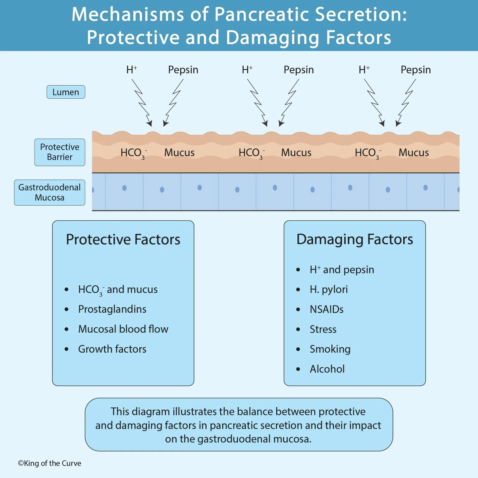
The stomach secretes hydrochloric acid (H⁺) and pepsin, both essential for digestion. However, these substances can also damage the mucosal lining if protective mechanisms are impaired.
The health of the gastroduodenal mucosa depends on the balance between:
Protective factors that preserve the mucosal barrier
Damaging factors that disrupt the barrier and promote ulcer formation
🧱 Protective Barrier of the Gastroduodenal Mucosa
The first line of defense is the mucus-bicarbonate barrier, which coats the epithelial surface.
🟤 Mucus
Surface epithelial cells secrete mucus that forms a thick gel layer over the mucosa. This layer:
Physically separates acid from epithelial cells
Traps bicarbonate ions
Slows diffusion of hydrogen ions
Protects against pepsin digestion
⚗️ Bicarbonate (HCO₃⁻)
Bicarbonate is secreted into the mucus layer where it neutralizes hydrogen ions.
This creates a nearly neutral pH immediately adjacent to epithelial cells despite the acidic gastric lumen.
Its functions include:
Neutralizing gastric acid
Maintaining epithelial cell viability
Preventing acid penetration
🛡️ Additional Protective Factors
💊 1. Prostaglandins
Prostaglandins (particularly PGE₂ and PGI₂) are among the most important endogenous protective factors.
They:
Increase mucus secretion
Increase bicarbonate secretion
Enhance mucosal blood flow
Promote epithelial repair
Reduce gastric acid secretion
NSAIDs impair these protective effects by inhibiting prostaglandin synthesis.
❤️ 2. Mucosal Blood Flow
Adequate blood flow supplies:
Oxygen
Nutrients
Bicarbonate
It also removes:
Diffused hydrogen ions
Toxic metabolites
Reduced perfusion increases susceptibility to ischemic injury and ulceration.
🌱 3. Growth Factors
Growth factors promote:
Rapid epithelial regeneration
Cell migration
Tissue repair
Restoration of the mucosal barrier following injury
This explains why superficial erosions often heal rapidly under normal conditions.
⚠️ Damaging Factors
When damaging influences overwhelm protective mechanisms, mucosal injury develops.
🧪 1. Hydrochloric Acid (H⁺)
Hydrogen ions are essential for digestion but can diffuse into damaged mucosa.
Excess acid:
Injures epithelial cells
Promotes inflammation
Delays ulcer healing
🔬 2. Pepsin
Pepsin is a proteolytic enzyme activated in acidic conditions.
It contributes to tissue injury by:
Digesting exposed proteins
Worsening existing mucosal defects
Preventing healing of ulcers
Acid and pepsin act together to produce significant mucosal damage.
🦠 3. Helicobacter pylori
H. pylori is the most common cause of peptic ulcer disease worldwide.
It causes injury by:
Producing urease
Triggering chronic inflammation
Damaging epithelial cells
Weakening the mucus layer
Increasing gastrin secretion in some patients
Persistent infection significantly increases ulcer risk.
💉 4. NSAIDs
Nonsteroidal anti-inflammatory drugs inhibit cyclooxygenase (COX), reducing prostaglandin production.
This results in:
Reduced mucus secretion
Reduced bicarbonate secretion
Decreased mucosal blood flow
Increased acid-mediated injury
Common NSAIDs include:
Ibuprofen
Naproxen
Diclofenac
Aspirin
😰 5. Physiological Stress
Severe illness can impair mucosal defenses through:
Reduced blood flow
Ischemia
Increased acid production
Stress ulcers commonly occur in:
ICU patients
Burn victims
Major trauma
Sepsis
🚬 6. Smoking
Smoking contributes to ulcer disease by:
Reducing mucosal blood flow
Decreasing bicarbonate secretion
Delaying ulcer healing
Increasing recurrence rates
Smoking cessation significantly improves healing.
🍺 7. Alcohol
Alcohol directly irritates the gastric mucosa.
Excessive intake may:
Increase epithelial permeability
Promote inflammation
Impair repair mechanisms
Exacerbate existing ulcers
🩺 Clinical Significance
Peptic ulcer disease develops when protective mechanisms fail to counteract damaging influences.
Common causes include:
H. pylori infection
Chronic NSAID use
Excess gastric acid secretion
Smoking
Severe physiological stress
Treatment aims to restore the balance by:
Eradicating H. pylori
Reducing acid secretion with proton pump inhibitors (PPIs)
Avoiding NSAIDs when possible
Encouraging smoking and alcohol cessation
Supporting mucosal healing
📋 High-Yield Summary
| 🛡️ Protective Factors | ⚠️ Damaging Factors |
|---|---|
| Mucus | Hydrochloric acid (H+) |
| Bicarbonate (HCO3−) | Pepsin |
| Prostaglandins | Helicobacter pylori |
| Adequate mucosal blood flow | NSAIDs |
| Growth factors | Physiological stress |
| Rapid epithelial regeneration | Smoking |
| Tight epithelial junctions | Alcohol |
🎯 Key Takeaway
The gastroduodenal mucosa remains healthy through a delicate balance between protective mechanisms—such as mucus, bicarbonate, prostaglandins, blood flow, and growth factors—and damaging influences like gastric acid, pepsin, Helicobacter pylori, NSAIDs, smoking, alcohol, and physiological stress. Disruption of this balance leads to mucosal injury and peptic ulcer disease, making restoration of mucosal protection a central goal of treatment.

Frequently Asked Questions (FAQs)
-
Aim for 4-6 focused hours, ensuring you incorporate breaks to avoid burnout.
-
Practice mindfulness techniques, take practice exams under realistic conditions, and maintain a balanced lifestyle.
-
Set short-term goals, seek support from mentors, and reward yourself for small achievements.
-
Regular exercise improves focus, reduces stress, and enhances overall mental clarity.
-
KOTC offers personalized learning tools, gamification features, and adaptive question banks to help students stay on track without burnout.