
💉 Understanding Lower GI Bleeding
Lower gastrointestinal (GI) bleeding is defined as bleeding originating distal to the ligament of Treitz, typically from the colon, rectum, or anus. It presents most commonly as hematochezia—bright red or maroon-colored blood in the stool—and can range from minor bleeding to massive hemorrhage. Prompt identification of the bleeding source is critical for stabilizing the patient and preventing complications.
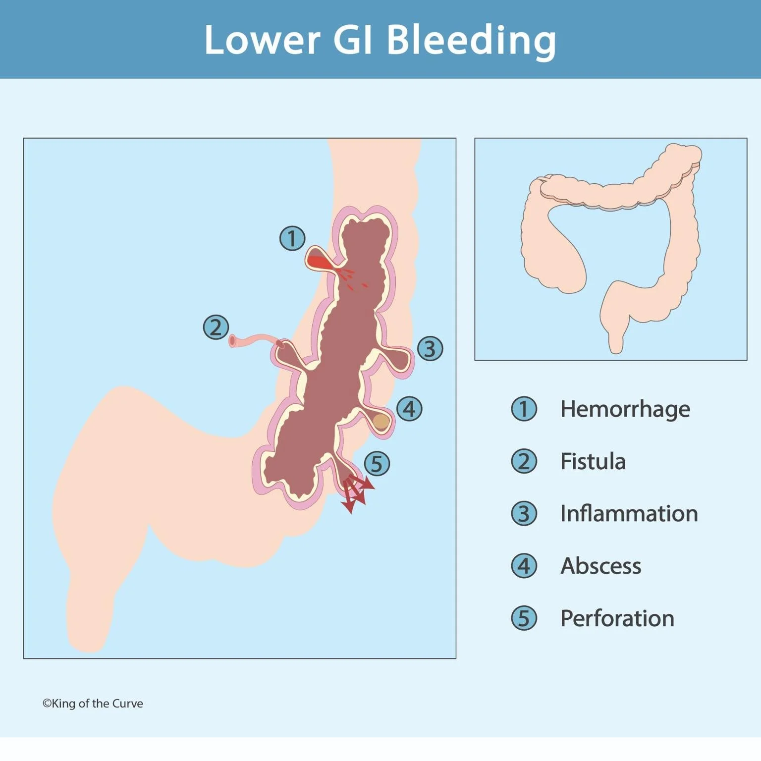
🩸 Common Causes of Lower GI Bleeding
The illustration highlights five key pathological mechanisms responsible for lower GI bleeding:
Hemorrhage (①) – Usually due to diverticulosis or angiodysplasia, where fragile vessels rupture under pressure.
Fistula (②) – An abnormal passage, such as a colovesical fistula, often seen in Crohn’s disease or post-surgical complications.
Inflammation (③) – Caused by ulcerative colitis, infectious colitis, or ischemic colitis leading to mucosal erosion.
Abscess (④) – A localized infection that can erode blood vessels and cause both pain and bleeding.
Perforation (⑤) – A full-thickness rupture of the intestinal wall, which can lead to peritonitis and sepsis—an emergency condition.
🧠 Diagnosis and Management
Lower GI bleeding is typically evaluated using colonoscopy, which serves both diagnostic and therapeutic purposes. In unstable patients, CT angiography helps identify active bleeding sites. Treatment may involve endoscopic hemostasis, embolization, or surgical resection, depending on severity. Supportive care includes IV fluids, blood transfusions, and monitoring hemodynamic stability.
⚖️ Upper vs. Lower GI Bleeding – Comparison Table
| Feature | Upper GI Bleed | Lower GI Bleed |
|---|---|---|
| Anatomical Source | Esophagus, stomach, duodenum | Colon, rectum, anus |
| Appearance of Stool | Melena (black, tarry stool) | Hematochezia (bright red or maroon) |
| Common Causes | Peptic ulcers, varices, gastritis | Diverticulosis, hemorrhoids, IBD |
| Diagnostic Tool | Upper endoscopy (EGD) | Colonoscopy or CT angiography |
| Treatment Approach | Proton pump inhibitors, endoscopic banding | Endoscopic cauterization; surgery if severe |
🧬 Complications and Prognosis
Untreated lower GI bleeding can lead to chronic anemia, hemodynamic shock, or infection in cases involving abscesses or perforations. The prognosis depends on the underlying cause and timeliness of intervention. Early diagnosis not only improves patient outcomes but also prevents recurrence through appropriate management and follow-up.
📚 Clinical Insight
In exams and real-life scenarios, differentiating upper vs. lower GI bleeding is essential. Remember: bright red blood = distal source, while dark, tarry stool = proximal source. Clinicians must also consider mixed presentations in cases of massive upper GI bleeds that rapidly transit through the intestines.
🚀 Call to Action – Master Medicine with King of the Curve
At King of the Curve, we make complex medical concepts simple, visual, and unforgettable. From anatomy to pathology, our high-yield illustrations and quizzes help students prepare smarter for exams like the MCAT, USMLE, and NCLEX.

Frequently Asked Questions (FAQs)
-
Aim for 4-6 focused hours, ensuring you incorporate breaks to avoid burnout.
-
Practice mindfulness techniques, take practice exams under realistic conditions, and maintain a balanced lifestyle.
-
Set short-term goals, seek support from mentors, and reward yourself for small achievements.
-
Regular exercise improves focus, reduces stress, and enhances overall mental clarity.
-
KOTC offers personalized learning tools, gamification features, and adaptive question banks to help students stay on track without burnout.