
🦵 Understanding the Lower Limb Arteries: Pathways of Circulation
The lower limb arteries form an intricate network responsible for delivering oxygen-rich blood to the muscles and tissues of the legs. These arteries not only sustain movement and endurance but also play a crucial role in maintaining vascular health — an essential topic for anyone studying anatomy, physiology, or preparing for exams like the MCAT, NCLEX, or USMLE.
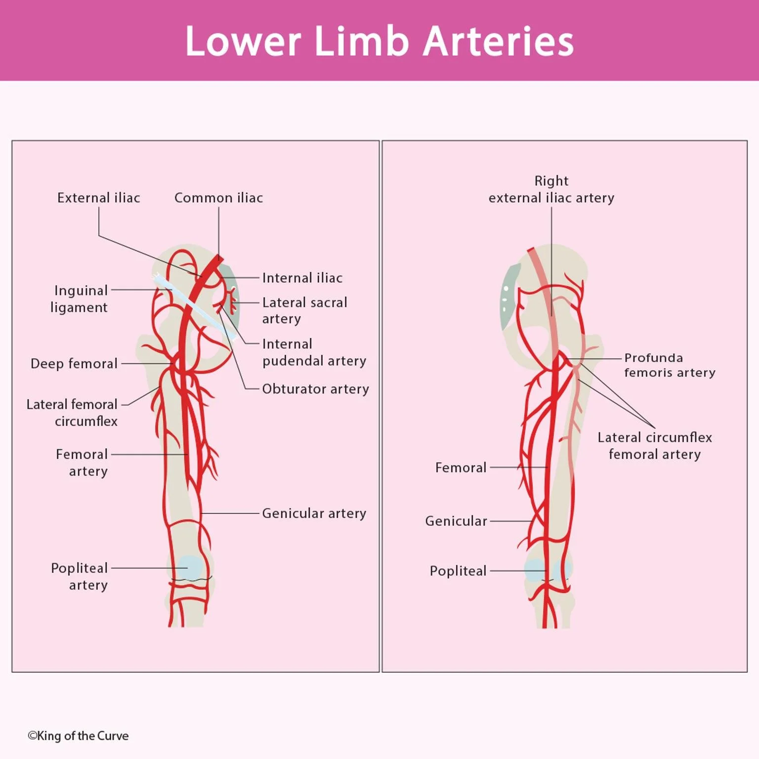
🩸 The Main Arterial Pathway
Blood supply to the lower limb begins from the common iliac artery, which divides into the internal and external iliac arteries.
The internal iliac artery provides branches such as the obturator artery, internal pudendal artery, and lateral sacral artery, supplying the pelvic region.
The external iliac artery, upon crossing the inguinal ligament, becomes the femoral artery — the major vessel responsible for lower limb perfusion.
As it travels down the thigh, the femoral artery gives rise to the deep femoral (profunda femoris) artery, which further branches into the lateral circumflex femoral artery and genicular arteries, supporting the thigh muscles and knee joint.
🦶 Below the Knee: Popliteal and Beyond
At the posterior aspect of the knee, the femoral artery transitions into the popliteal artery. This vessel divides into several smaller arteries that ensure proper blood distribution to the calf and foot.
The genicular arteries also contribute to collateral circulation around the knee, a vital adaptation for maintaining blood flow during bending or compression.
Understanding this flow pathway is particularly important in clinical practice — from evaluating peripheral artery disease (PAD) to managing traumatic limb injuries and performing vascular surgeries.
📘 Table: Key Arteries of the Lower Limb
| Region | Main Arteries | Clinical Significance |
|---|---|---|
| Pelvic Region | Common iliac, internal iliac, external iliac | Supplies pelvic organs and lower limbs |
| Thigh | Femoral, profunda femoris, lateral circumflex femoral | Common site for catheter insertion and pulse assessment |
| Knee | Genicular arteries | Important for collateral blood flow during flexion |
| Leg | Popliteal, anterior & posterior tibial arteries | Prone to obstruction in peripheral artery disease |
| Foot | Dorsalis pedis, plantar arteries | Checked for circulation and diabetic foot evaluation |
🧠 KOTC Tip for Exam Prep
When tackling MCAT or USMLE-style anatomy questions, visualize the arterial flow direction:
Common Iliac → External Iliac → Femoral → Popliteal → Tibial → Pedal Arteries.
Recognizing this order helps students quickly identify clinical implications such as claudication, embolism, or aneurysm formation.
🩺 Clinical Relevance: Diagnosing Vascular Compromise
Disorders of the lower limb arteries are commonly diagnosed through imaging modalities like Doppler ultrasound, CT angiography, or MR angiography. A reduced pulse in the dorsalis pedis or posterior tibial arteries may indicate arterial insufficiency. Understanding arterial branching allows clinicians to localize blockages and plan effective interventions such as angioplasty or bypass surgery. For students, remembering the hierarchy of arterial flow can also improve comprehension of pathology case studies and clinical correlations.
🚀 Call to Action
Master human anatomy with interactive visuals and adaptive quizzes on the King of the Curve App!
Our 3D illustrations, daily QOTDs, and KOTC Classroom make complex concepts — like the lower limb arterial system — effortless to learn and retain.

Frequently Asked Questions (FAQs)
-
Aim for 4-6 focused hours, ensuring you incorporate breaks to avoid burnout.
-
Practice mindfulness techniques, take practice exams under realistic conditions, and maintain a balanced lifestyle.
-
Set short-term goals, seek support from mentors, and reward yourself for small achievements.
-
Regular exercise improves focus, reduces stress, and enhances overall mental clarity.
-
KOTC offers personalized learning tools, gamification features, and adaptive question banks to help students stay on track without burnout.