
🧬 How PET Scans Work: A Molecular Level Breakdown
Positron Emission Tomography, or PET scanning, is one of the most advanced medical imaging technologies used to visualize activity at the molecular level. Unlike CT or MRI scans that focus on structure, PET scans reveal function and metabolism—how organs and tissues are actually working.
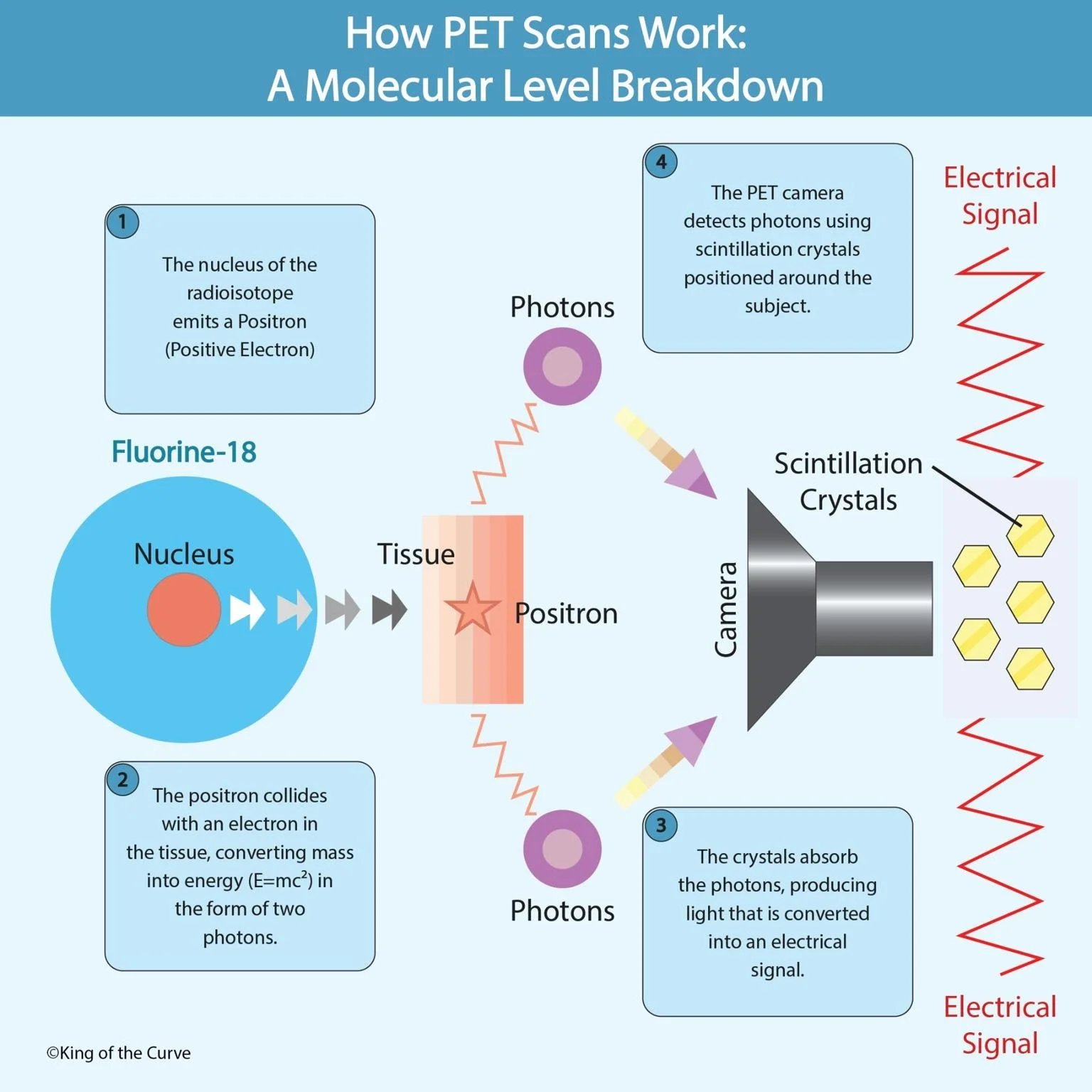
🧠 Step 1: The Radioisotope Emits a Positron
PET scans begin with the injection of a radioactive tracer, commonly Fluorine-18, into the patient’s body. This isotope emits a positron, which is a positively charged electron. The tracer is usually attached to glucose, so it travels to areas with high energy use—like the brain, heart, or cancerous tissues.
⚛️ Step 2: Collision and Annihilation
As the positron moves through the body, it soon collides with an electron. When these two particles meet, they annihilate each other—releasing energy in the form of two photons (light particles) that travel in opposite directions. This energy conversion follows Einstein’s equation E = mc².
💡 Step 3: Photon Detection
The photons created during annihilation are detected by a ring of scintillation crystals inside the PET scanner. These crystals convert the photon energy into light signals, which are then transformed into electrical signals by sensitive detectors.
🖥️ Step 4: Image Construction
Finally, a computer processes these electrical signals to create 3D images of the body’s metabolic activity. Regions with higher tracer concentration—indicating increased metabolic function—appear as bright spots on the PET scan.
🩺 What PET Scans Reveal
PET scans are used to:
Detect cancerous tumors and monitor treatment effectiveness.
Measure brain activity in neurological conditions like Alzheimer’s or epilepsy.
Assess heart muscle function after a heart attack or during stress testing.
By visualizing molecular activity, PET scans help doctors diagnose disease before physical changes occur—making them a crucial tool in early detection and precision medicine.
⚖️ PET vs. MRI vs. CT
| Feature | PET Scan | MRI Scan | CT Scan |
|---|---|---|---|
| Purpose | Shows metabolic & biochemical activity | Shows detailed soft-tissue structure | Shows structural anatomy |
| Uses Radiation? | ✅ Yes (radioisotopes) | ❌ No | ✅ Yes (X-rays) |
| Best For | Cancer detection Brain activity Metabolism | Brain & spine Joints Soft tissues | Bones Lungs Trauma |
| Type of Image | Functional | Anatomical | Anatomical |
| Time Required | 30–60 minutes | 30–90 minutes | 5–10 minutes |
| Cost | High 💰 | Moderate 💵 | Lower 💸 |
🧩 Summary
PET scans combine nuclear physics, molecular biology, and advanced imaging to provide a real-time view of the body’s inner workings. By tracking biochemical processes instead of just physical structures, PET technology gives clinicians an unmatched ability to understand disease progression and tailor treatments accordingly.
🧠 Quick Fact
The most common PET tracer, Fluorine-18, has a half-life of just under two hours—short enough to minimize radiation exposure, but long enough to complete high-quality imaging.
🧭 In a Nutshell
PET scans turn invisible molecular activity into visible diagnostic data. They don’t just show what’s inside the body—they show how it’s working, making them a vital tool in modern medicine.

Frequently Asked Questions (FAQs)
-
Aim for 4-6 focused hours, ensuring you incorporate breaks to avoid burnout.
-
Practice mindfulness techniques, take practice exams under realistic conditions, and maintain a balanced lifestyle.
-
Set short-term goals, seek support from mentors, and reward yourself for small achievements.
-
Regular exercise improves focus, reduces stress, and enhances overall mental clarity.
-
KOTC offers personalized learning tools, gamification features, and adaptive question banks to help students stay on track without burnout.