
🩸 Pathway of Heme Synthesis and Related Disorders
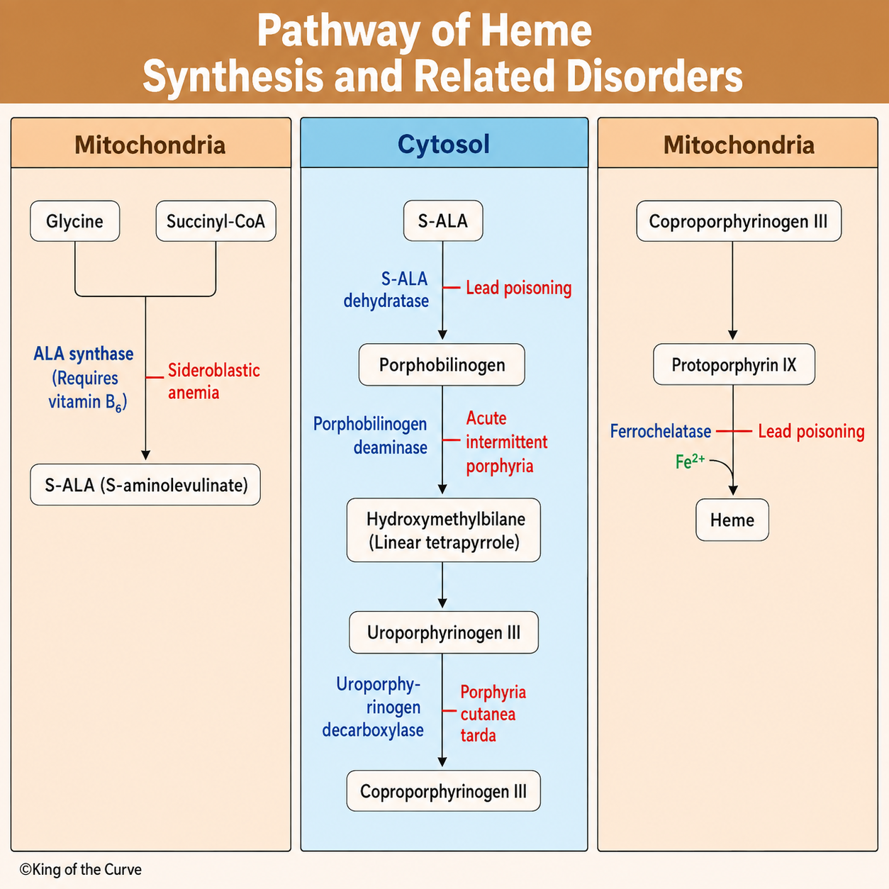
Heme synthesis is a fundamental biochemical pathway responsible for producing heme, the iron-containing molecule found in hemoglobin, myoglobin, cytochromes, and numerous enzymes involved in oxygen transport and cellular respiration. Because this pathway spans both the mitochondria and cytosol, defects in specific enzymes can lead to characteristic disorders known as porphyrias or sideroblastic anemia. Understanding the heme synthesis pathway and its associated diseases is highly tested on the MCAT and forms an essential foundation for medical education. The King of the Curve infographic provides a step-by-step visual guide to this important metabolic pathway.
Where Heme Synthesis Begins
Heme synthesis starts inside the mitochondria, where glycine combines with succinyl-CoA to form δ-aminolevulinic acid (ALA) through the enzyme ALA synthase. This first and rate-limiting step requires vitamin B₆ (pyridoxine) as a cofactor. Vitamin B₆ deficiency or impaired ALA synthase activity can result in sideroblastic anemia, a condition characterized by defective heme production despite adequate iron stores. Once ALA is formed, it moves into the cytosol for the next series of enzymatic reactions.
⚙️ The Cytosolic Steps of Heme Synthesis
Within the cytosol, ALA is converted into porphobilinogen by ALA dehydratase, an enzyme inhibited by lead poisoning. Porphobilinogen is then converted into hydroxymethylbilane through porphobilinogen deaminase, whose deficiency causes acute intermittent porphyria (AIP). The pathway continues through the formation of uroporphyrinogen III and coproporphyrinogen III, with uroporphyrinogen decarboxylase deficiency resulting in porphyria cutanea tarda (PCT), the most common porphyria.
⚠️ Lead Poisoning and Heme Synthesis
Lead poisoning disrupts heme production by inhibiting two critical enzymes: ALA dehydratase and ferrochelatase. As a result, ALA and protoporphyrin accumulate while heme production decreases. Clinically, patients may present with microcytic anemia, abdominal pain, peripheral neuropathy, developmental delay, and basophilic stippling of red blood cells. On the MCAT, remembering that lead inhibits both ALA dehydratase and ferrochelatase is one of the highest-yield facts in biochemistry.
📋 Key Enzymes and Related Disorders
| Enzyme | Function | Associated Disorder / Inhibitor |
|---|---|---|
| ALA Synthase | Forms ALA from glycine and succinyl-CoA. | Vitamin B6 deficiency → Sideroblastic anemia |
| ALA Dehydratase | Converts ALA to porphobilinogen. | Inhibited by lead poisoning |
| Porphobilinogen Deaminase | Produces hydroxymethylbilane. | Acute intermittent porphyria |
| Uroporphyrinogen Decarboxylase | Forms coproporphyrinogen III. | Porphyria cutanea tarda |
| Ferrochelatase | Inserts Fe2+ into protoporphyrin IX. | Inhibited by lead poisoning |
🩺 Clinical Significance
Disorders of heme synthesis produce distinctive clinical presentations. Acute intermittent porphyria typically presents with severe abdominal pain, psychiatric symptoms, and peripheral neuropathy without photosensitivity. In contrast, porphyria cutanea tarda causes blistering skin lesions and photosensitivity due to the accumulation of light-sensitive porphyrins. Sideroblastic anemia results from impaired heme production, while lead poisoning produces anemia along with neurological and gastrointestinal symptoms. Recognizing these disease patterns is essential for both clinical practice and standardized exams.
📚 Why This Topic Is High-Yield for the MCAT
The MCAT frequently integrates heme synthesis with biochemistry, metabolism, genetics, hematology, and toxicology. Students may be asked to identify the enzyme affected by lead poisoning, determine which vitamin deficiency causes sideroblastic anemia, or recognize the symptoms of various porphyrias. A useful memory aid is: Lead blocks ALA Dehydratase and Ferrochelatase, Vitamin B₆ is required for ALA Synthase, Acute Intermittent Porphyria involves Porphobilinogen Deaminase, and Porphyria Cutanea Tarda involves Uroporphyrinogen Decarboxylase.
🎯 Key Takeaway
The heme synthesis pathway spans the mitochondria and cytosol, requiring multiple enzymes to convert glycine and succinyl-CoA into heme. Defects in these enzymes produce characteristic disorders, including sideroblastic anemia, acute intermittent porphyria, porphyria cutanea tarda, and lead poisoning. By understanding the sequence of enzymatic reactions, associated cofactors, and disease presentations, you'll be well prepared to answer high-yield MCAT questions involving metabolism, hematology, and biochemical pathways.

Frequently Asked Questions (FAQs)
-
Aim for 4-6 focused hours, ensuring you incorporate breaks to avoid burnout.
-
Practice mindfulness techniques, take practice exams under realistic conditions, and maintain a balanced lifestyle.
-
Set short-term goals, seek support from mentors, and reward yourself for small achievements.
-
Regular exercise improves focus, reduces stress, and enhances overall mental clarity.
-
KOTC offers personalized learning tools, gamification features, and adaptive question banks to help students stay on track without burnout.