
🫁 Esophageal Atresia and Tracheoesophageal Fistula
Esophageal atresia (EA) is a congenital abnormality in which the esophagus does not form a continuous passage from the pharynx to the stomach. Tracheoesophageal fistula (TEF) is an abnormal connection between the trachea and esophagus. These defects arise during embryologic development and are often discussed together because they commonly occur in combination.
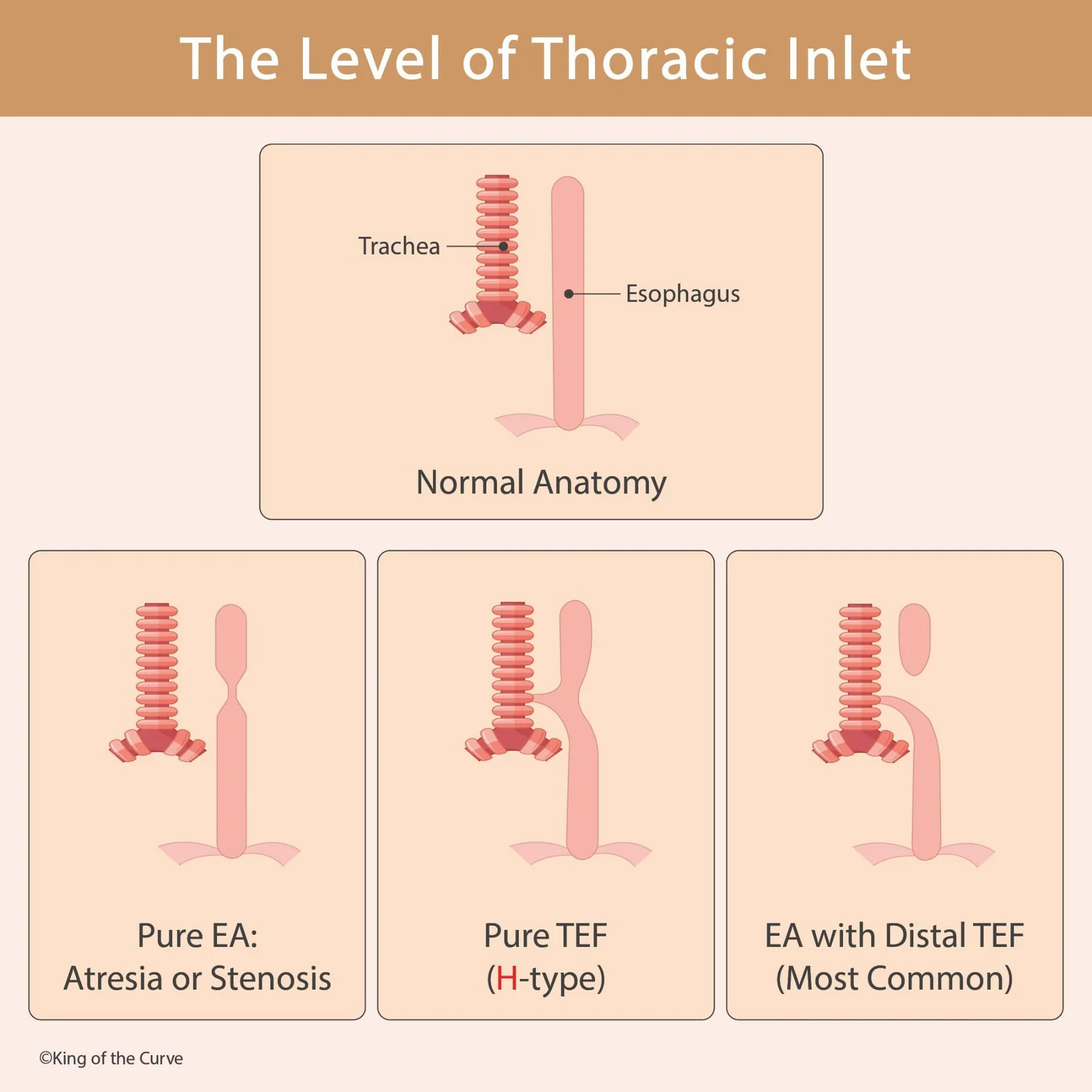
🫁 Normal Trachea and Esophagus Anatomy
Normally, the trachea lies anterior to the esophagus. During embryonic development, the foregut separates into a ventral respiratory tract and a dorsal esophagus. Proper separation allows swallowed material to enter the gastrointestinal tract while inhaled air travels through the respiratory tract.
🧫 How Do EA and TEF Develop?
EA and TEF are associated with abnormal separation of the primitive foregut into the trachea and esophagus. Problems during this developmental process can leave the esophagus discontinuous, create an abnormal fistulous connection with the trachea, or produce both abnormalities.
🔬 Major Anatomical Patterns
Several anatomical configurations are possible. Pure esophageal atresia involves an interrupted esophagus without a tracheoesophageal fistula. H-type TEF consists of an abnormal tracheoesophageal connection without esophageal atresia. The most common configuration is esophageal atresia with a distal TEF, in which the upper esophagus ends blindly while the distal esophageal segment communicates with the trachea.
| 🫁 Pattern | 📘 Esophagus | 🔗 Tracheoesophageal Connection |
|---|---|---|
| Pure EA | Discontinuous | Absent |
| H-type TEF | Continuous | Present |
| EA with distal TEF | Discontinuous | Distal fistula present |
👶 Common Clinical Presentation
Newborns with esophageal atresia may develop excessive salivation, coughing, choking, or cyanosis during attempted feeding. Because the esophagus is interrupted, feeds cannot pass normally into the stomach. Aspiration can also occur, particularly when a fistulous connection allows material to enter the respiratory tract.
🩻 How Is Esophageal Atresia Detected?
A classic clue is difficulty passing a nasogastric or orogastric tube into the stomach. Imaging may demonstrate the tube coiled within the proximal esophageal pouch. The presence or absence of gas in the stomach and intestines can also provide information about whether a distal TEF is present.
🤰 Prenatal Clues
Esophageal atresia can interfere with fetal swallowing and may therefore be associated with polyhydramnios during pregnancy. Prenatal ultrasound may raise suspicion, although definitive characterization of the anatomy generally requires evaluation after birth.
🧠 Associated Congenital Abnormalities
EA and TEF can occur alongside other congenital abnormalities. A commonly tested association is VACTERL, which includes vertebral, anorectal, cardiac, tracheoesophageal, renal, and limb abnormalities. Recognizing this association can help connect embryology with broader congenital-development concepts.
🎯 High-Yield Takeaway
For exam review, remember the basic distinction: EA means the esophagus is interrupted, while TEF means there is an abnormal connection between the trachea and esophagus. EA with distal TEF is the most common configuration. Key clues include feeding-related choking, excessive secretions, inability to pass a feeding tube, and possible polyhydramnios before birth.

Frequently Asked Questions (FAQs)
-
Aim for 4-6 focused hours, ensuring you incorporate breaks to avoid burnout.
-
Practice mindfulness techniques, take practice exams under realistic conditions, and maintain a balanced lifestyle.
-
Set short-term goals, seek support from mentors, and reward yourself for small achievements.
-
Regular exercise improves focus, reduces stress, and enhances overall mental clarity.
-
KOTC offers personalized learning tools, gamification features, and adaptive question banks to help students stay on track without burnout.