
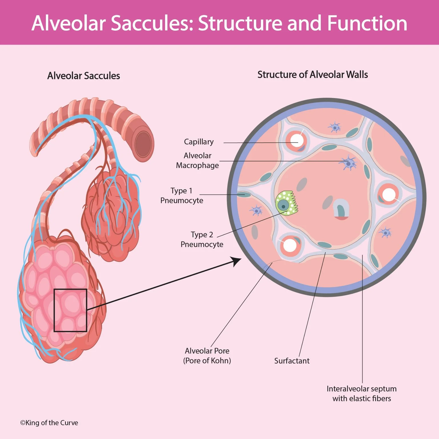
Alveolar sacs are clusters of tiny air spaces called alveoli at the ends of the lung’s respiratory passages. Each alveolus provides a surface where air comes into close contact with pulmonary capillary blood. This arrangement allows oxygen to enter the bloodstream and carbon dioxide to leave it during breathing.
💨 How Gases Move Between Air and Blood
Oxygen and carbon dioxide cross the alveolar–capillary barrier by passive diffusion, following their respective partial pressure gradients. Oxygen normally moves from alveolar air into incoming pulmonary capillary blood, while carbon dioxide moves in the opposite direction. Effective exchange depends on both ventilation, which supplies fresh air, and perfusion, which supplies blood.
🧱 Why the Air–Blood Barrier Is Thin
The thin portion of the air–blood barrier includes a type I pneumocyte, closely associated basement membranes, and a capillary endothelial cell. Its short diffusion distance helps gases move efficiently between alveolar air and blood. A large exchange surface further supports this process, while increased barrier thickness can slow diffusion.
🫧 Type I Pneumocytes: Built for Gas Exchange
Type I pneumocytes are broad, flattened epithelial cells that cover approximately 95% of the alveolar surface. Their thin shape makes them well suited for gas exchange without creating a thick barrier. They also help maintain the integrity of the alveolar lining, separating the air spaces from the underlying tissue and circulation.
📊 Key Alveolar Structures and Their Functions
Alveoli rely on several specialized cells and supporting structures rather than a single cell type. Some provide a thin exchange surface, others produce surfactant or remove inhaled material, and connective tissue supports expansion and recoil. The table summarizes how these components work together.
| Layer | Location | Main Hormone Products | Main Regulation | Major Role |
|---|---|---|---|---|
| Zona glomerulosa | Outermost cortex | Aldosterone | Angiotensin II and potassium | Sodium balance, potassium excretion, and blood pressure |
| Zona fasciculata | Middle cortex | Cortisol | ACTH | Metabolism and stress responses |
| Zona reticularis | Innermost cortex | DHEA, DHEA-S, and androstenedione | ACTH | Production of androgen precursors |
🧪 Type II Pneumocytes: Surfactant and Repair
Type II pneumocytes produce pulmonary surfactant and store its components in structures called lamellar bodies. Surfactant reduces surface tension at the alveolar air–liquid interface, helping stabilize alveoli and reduce their tendency to collapse during expiration. Type II cells also contribute to repair by proliferating and replacing damaged alveolar epithelial cells.
🛡️ Alveolar Macrophages: Local Immune Defense
Alveolar macrophages, sometimes called dust cells, patrol the alveolar environment and engulf inhaled particles, microorganisms, and cellular debris. This phagocytic activity helps protect the delicate gas exchange surfaces. They also participate in immune signaling, helping coordinate local responses when potentially harmful material reaches the alveoli.
🔗 Pores of Kohn and Elastic Support
Pores of Kohn are small openings connecting neighboring alveoli, allowing collateral airflow and helping equalize pressure between them. The interalveolar septa contain capillaries and supporting connective tissue, including elastic fibers. These fibers stretch during inspiration and contribute to passive recoil during quiet expiration.
⚠️ How Structural Damage Impairs Gas Exchange
Gas exchange becomes less efficient when alveolar surface area decreases or the diffusion barrier thickens. Emphysema reduces the available exchange surface through destruction of alveolar walls, while pulmonary fibrosis increases diffusion distance through scarring. These examples show why healthy lungs need both an extensive alveolar surface and a thin, intact air–blood barrier.

Frequently Asked Questions (FAQs)
-
Aim for 4-6 focused hours, ensuring you incorporate breaks to avoid burnout.
-
Practice mindfulness techniques, take practice exams under realistic conditions, and maintain a balanced lifestyle.
-
Set short-term goals, seek support from mentors, and reward yourself for small achievements.
-
Regular exercise improves focus, reduces stress, and enhances overall mental clarity.
-
KOTC offers personalized learning tools, gamification features, and adaptive question banks to help students stay on track without burnout.