
❤️ Spike and Dome Pulse Curve: Understanding Pulsus Bisferiens
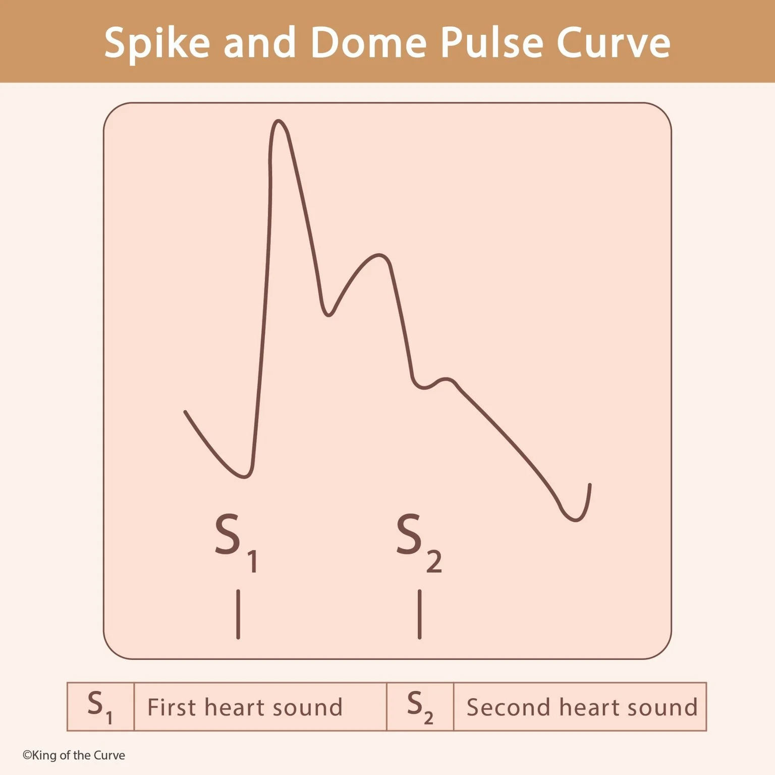
A spike and dome pulse is an arterial pulse waveform characterized by two systolic peaks. The first is a sharp, rapidly rising spike, followed by a brief decline and then a second, broader dome. This pattern is classically associated with hypertrophic obstructive cardiomyopathy (HOCM) and can also be seen in some patients with combined aortic stenosis and aortic regurgitation.
📈 Why Does the Pulse Have Two Peaks?
In HOCM, the left ventricle initially ejects blood rapidly, producing the first prominent systolic peak. As ventricular contraction continues, dynamic narrowing of the left ventricular outflow tract (LVOT) can increase, temporarily reducing forward blood flow. Later changes in ventricular ejection produce the second, more rounded portion of the waveform, giving the pulse its characteristic spike-and-dome appearance.
🫀 The Connection to Hypertrophic Obstructive Cardiomyopathy
HOCM is characterized by abnormal thickening of the ventricular myocardium, often involving the interventricular septum. During systole, the narrowed outflow tract and systolic anterior motion of the mitral valve can dynamically obstruct blood leaving the left ventricle. Because the degree of obstruction can change with ventricular volume and contractility, HOCM produces several distinctive findings that are important for cardiovascular physiology questions.
🔊 Understanding S1 and S2
The diagram also marks the first and second heart sounds. S1 occurs primarily when the mitral and tricuspid valves close at the beginning of ventricular systole. S2 occurs when the aortic and pulmonary valves close near the end of systole. The arterial pulse waveform between these sounds therefore largely corresponds to ventricular ejection during systole.
🩺 Pulsus Bisferiens
A pulse with two palpable systolic peaks is called pulsus bisferiens. It can be appreciated in arteries such as the carotid artery and is associated particularly with HOCM and significant aortic regurgitation, sometimes in combination with aortic stenosis. Recognizing the double systolic peak can help connect a physical examination finding with the underlying cardiovascular physiology.
⚖️ Preload and the HOCM Murmur
HOCM is especially important because its dynamic obstruction changes with loading conditions. Decreasing preload makes the ventricular chamber smaller, bringing the hypertrophied septum and mitral apparatus closer together and increasing LVOT obstruction. As a result, maneuvers such as standing or the Valsalva maneuver generally make the HOCM murmur louder.
| ❤️ Feature | 🫀 HOCM Finding |
|---|---|
| Pulse pattern | Spike and dome / pulsus bisferiens |
| Primary problem | Dynamic LV outflow obstruction |
| Decreased preload | Increases obstruction |
| Valsalva | Murmur becomes louder |
| Squatting | Murmur generally becomes softer |
🧎 Why Squatting Changes the Murmur
Squatting generally increases venous return and systemic vascular resistance. The resulting increase in ventricular filling enlarges the ventricular cavity and tends to reduce the dynamic obstruction in HOCM. Consequently, the HOCM murmur usually decreases with squatting. This contrasts with standing or Valsalva, which reduce ventricular filling and typically increase the murmur.
🧠 Spike and Dome vs Other Pulse Findings
Do not confuse a spike-and-dome waveform with other arterial pulse abnormalities. Pulsus paradoxus refers to an exaggerated inspiratory decrease in systolic blood pressure and is classically associated with cardiac tamponade. Pulsus alternans involves alternating strong and weak beats and can indicate severe left ventricular dysfunction. Pulsus bisferiens specifically describes two systolic peaks within a single arterial pulse.
🎯 MCAT High-Yield Takeaway
For cardiovascular physiology, connect spike and dome → pulsus bisferiens → HOCM while remembering that pulsus bisferiens can occur in other valvular conditions as well. In HOCM, reduced ventricular filling increases dynamic LVOT obstruction, so Valsalva/standing makes the murmur louder, while squatting generally makes it softer. Also remember the timing of the heart sounds: S1 marks AV valve closure and the beginning of systole, while S2 marks semilunar valve closure and the end of systole.

Frequently Asked Questions (FAQs)
-
Aim for 4-6 focused hours, ensuring you incorporate breaks to avoid burnout.
-
Practice mindfulness techniques, take practice exams under realistic conditions, and maintain a balanced lifestyle.
-
Set short-term goals, seek support from mentors, and reward yourself for small achievements.
-
Regular exercise improves focus, reduces stress, and enhances overall mental clarity.
-
KOTC offers personalized learning tools, gamification features, and adaptive question banks to help students stay on track without burnout.