
🩺 Pancreatic Cancer Diagnosis Flow
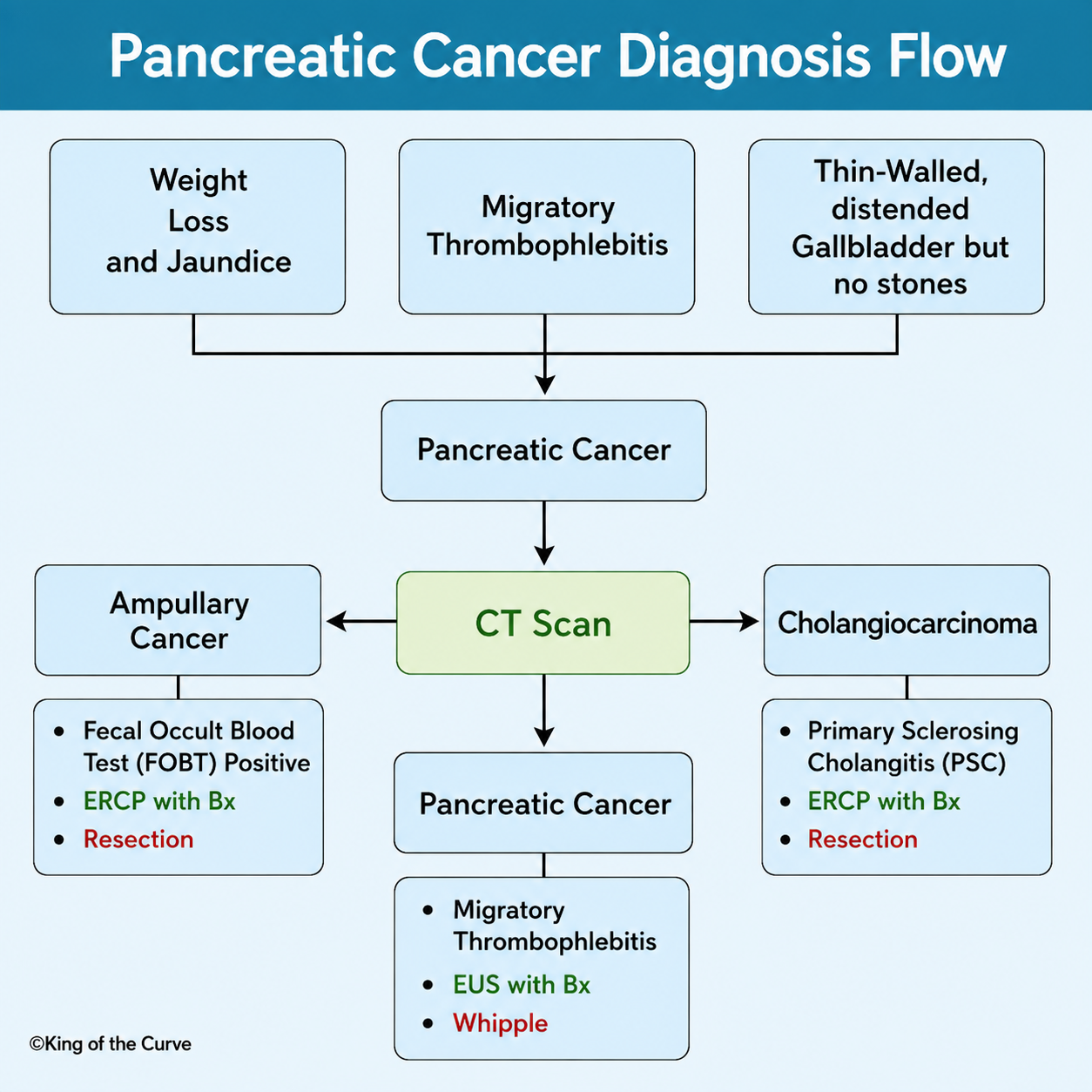
Pancreatic cancer is one of the deadliest malignancies because it often develops silently and is diagnosed only after it has reached an advanced stage. For students preparing for the MCAT, understanding the diagnostic approach is essential since pancreatic cancer frequently appears in passage-based questions involving gastrointestinal disease, oncology, and clinical reasoning. The King of the Curve diagnostic flow image simplifies this complex process into an easy-to-follow pathway that helps students recognize key clinical clues and make the correct diagnosis more efficiently.
⚠️ Recognizing the Classic Warning Signs
Patients with pancreatic cancer commonly present with unexplained weight loss, painless jaundice, and abdominal discomfort that may radiate to the back. Another classic finding is migratory thrombophlebitis (Trousseau syndrome), a condition caused by the cancer creating a hypercoagulable state. A thin-walled, distended gallbladder without gallstones, known as Courvoisier sign, is another hallmark feature suggesting obstruction by a pancreatic head tumor rather than gallstones. Recognizing these warning signs early is critical for both clinical practice and standardized exams.
🖥️ CT Scan: The First Diagnostic Step
Once pancreatic cancer is suspected, a contrast-enhanced CT scan is usually the first imaging study performed. CT imaging helps determine the location and size of the tumor, whether nearby blood vessels are involved, and if the cancer has spread to distant organs. The scan also helps distinguish pancreatic cancer from other conditions such as ampullary cancer and cholangiocarcinoma, both of which can produce similar symptoms. Accurate imaging guides physicians toward the most appropriate next diagnostic and treatment steps.
🔬 Confirming the Diagnosis
Although CT scans provide valuable information, a definitive diagnosis requires a tissue sample. The preferred method is Endoscopic Ultrasound (EUS) with biopsy, which allows physicians to visualize the pancreas closely while obtaining tissue for microscopic examination. This technique is highly accurate and minimally invasive, making it the gold standard for confirming pancreatic cancer before treatment planning begins. Histological confirmation is especially important when surgery or chemotherapy is being considered.
⚖️ Differential Diagnosis at a Glance
The following table summarizes the key conditions that should be differentiated when evaluating a patient with obstructive jaundice and suspected pancreatic disease.
| Condition | Key Clinical Clue | Diagnostic Test | Typical Treatment |
|---|---|---|---|
| Pancreatic Cancer | Weight loss, painless jaundice, and migratory thrombophlebitis | CT scan followed by EUS with biopsy | Whipple procedure if the tumor is resectable |
| Ampullary Cancer | Positive fecal occult blood test and obstructive jaundice | ERCP with biopsy | Surgical resection |
| Cholangiocarcinoma | Strong association with Primary Sclerosing Cholangitis | ERCP with biopsy | Surgical resection when possible |
🏥 Treatment and the Whipple Procedure
For patients with localized pancreatic cancer, surgery offers the best chance of long-term survival. The standard operation is the Whipple procedure (pancreaticoduodenectomy), which removes the head of the pancreas along with portions of the duodenum, bile duct, gallbladder, and sometimes part of the stomach. While this is one of the most complex abdominal surgeries, it remains the primary curative option for tumors that have not spread beyond the pancreas.
📚 Why This Matters for the MCAT
Pancreatic cancer is considered a high-yield MCAT topic because it combines concepts from pathology, anatomy, physiology, oncology, and diagnostic imaging. Exam passages often describe a patient with painless jaundice, weight loss, new-onset diabetes, or recurrent blood clots, expecting students to identify pancreatic adenocarcinoma and select the next appropriate diagnostic test. Memorizing these classic associations using visual learning tools can dramatically improve recall during the exam.
🎯 Key Takeaway
Pancreatic cancer is a high-yield disease for the MCAT because it integrates anatomy, pathology, physiology, oncology, and clinical reasoning into one diagnosis. Remember the classic triad of weight loss, painless jaundice, and Courvoisier sign, along with migratory thrombophlebitis (Trousseau syndrome) as important diagnostic clues. The standard diagnostic pathway begins with a contrast-enhanced CT scan, followed by Endoscopic Ultrasound (EUS) with biopsy for confirmation, while the Whipple procedure remains the primary curative treatment for eligible patients. Using visual study tools like the King of the Curve Pancreatic Cancer Diagnosis Flow can help reinforce these concepts, making them easier to recall on exam day and in future clinical practice.

Frequently Asked Questions (FAQs)
-
Aim for 4-6 focused hours, ensuring you incorporate breaks to avoid burnout.
-
Practice mindfulness techniques, take practice exams under realistic conditions, and maintain a balanced lifestyle.
-
Set short-term goals, seek support from mentors, and reward yourself for small achievements.
-
Regular exercise improves focus, reduces stress, and enhances overall mental clarity.
-
KOTC offers personalized learning tools, gamification features, and adaptive question banks to help students stay on track without burnout.