
🫀 Ligamentum Teres Hepatis: Anatomy, Function & Clinical Significance
The Ligamentum Teres Hepatis, also known as the round ligament of the liver, is a small but fascinating remnant of fetal circulation that continues to hold clinical importance in adult anatomy.
Understanding this ligament’s origin, location, and connections is essential for medical students, future surgeons, and anyone preparing for the MCAT, USMLE, or NCLEX.
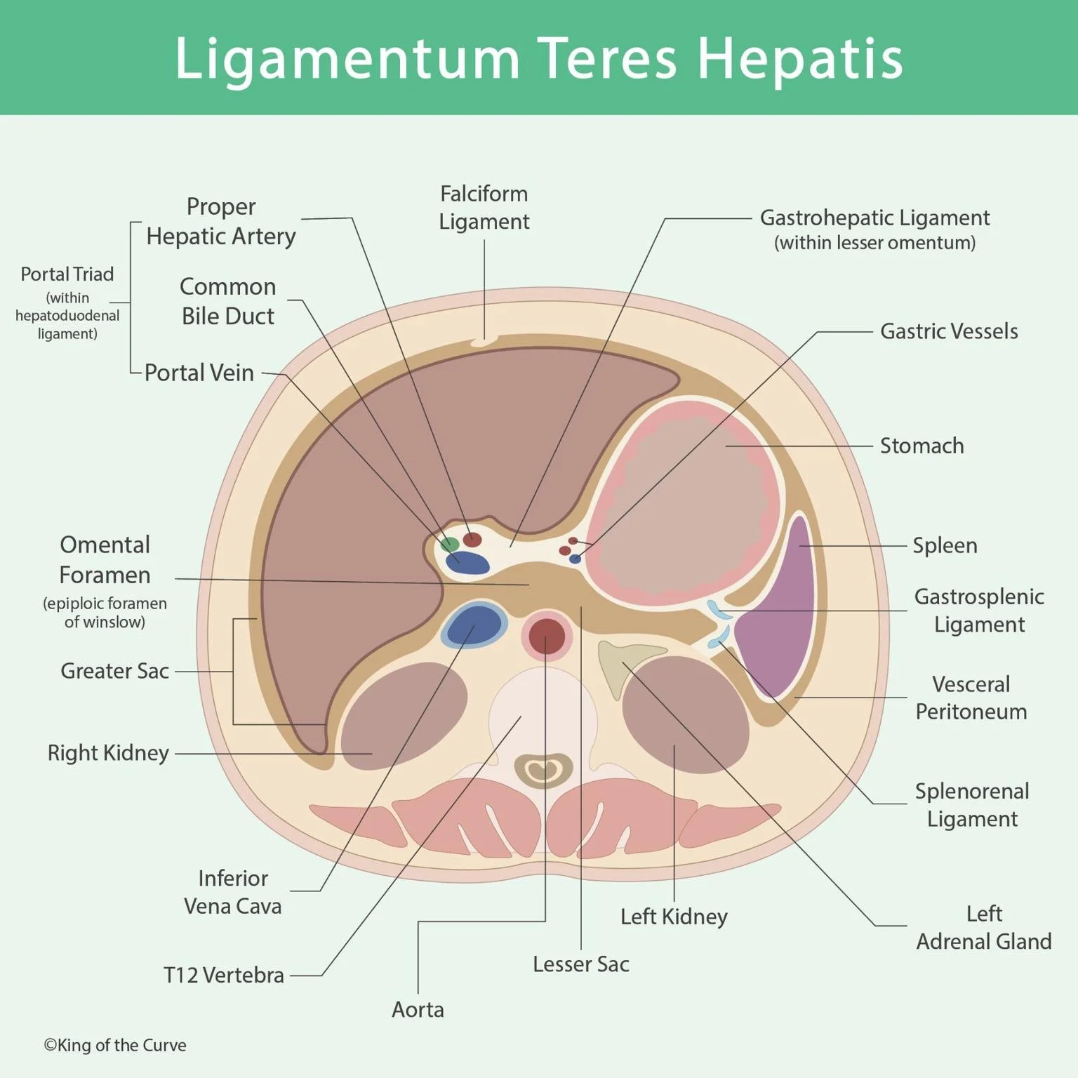
⚙️ Anatomy and Location
The Ligamentum Teres Hepatis is located within the inferior margin of the falciform ligament, which connects the liver to the anterior abdominal wall.
It runs from the umbilicus to the liver’s inferior surface, where it merges with the left branch of the portal vein.
The ligament is a fibrous remnant of the fetal umbilical vein, which once carried oxygenated blood from the placenta to the fetus.
🧬 Embryological Origin
During fetal life, the umbilical vein plays a crucial role in shunting oxygen-rich blood toward the developing fetus.
After birth:
The umbilical vein closes and fibroses, becoming the Ligamentum Teres Hepatis.
The closure of the ductus venosus, another fetal vessel, leads to the formation of the Ligamentum Venosum.
💡 MCAT Tip:
Remember — both the Ligamentum Teres Hepatis and Ligamentum Venosum are remnants of fetal circulation, making them great examples of developmental anatomy tested on the MCAT and USMLE.
📊 Quick Summary Table
| Feature | Description | Clinical Relevance |
|---|---|---|
| Structure | Fibrous remnant of the fetal umbilical vein | Located in the inferior border of the falciform ligament |
| Function (Fetal) | Carries oxygenated blood from placenta to fetus | Essential for fetal oxygen supply |
| Function (Adult) | No functional role; anatomical landmark | Separates left and right lobes of the liver |
| Associated Ligament | Falciform ligament | Connects liver to anterior abdominal wall |
| Clinical Significance | Recanalization in portal hypertension | Seen as “caput medusae” on physical exam |
💡 Exam Connection:
In portal hypertension, the Ligamentum Teres Hepatis can recanalize, allowing blood to bypass the liver via the paraumbilical veins — leading to distended abdominal veins (caput medusae).!
⚕️ Clinical Importance
1️⃣ Portal Hypertension:
The ligament can reopen, forming collateral pathways between the portal and systemic venous systems.
2️⃣ Surgical Landmark:
Surgeons use it to identify the falciform ligament and divide the liver lobes during procedures like hepatic resections.
3️⃣ Radiologic Marker:
Visible on ultrasound or CT scans as a linear echogenic band extending from the umbilicus to the liver — helpful in liver imaging interpretation.
🧩 Exam Integration
MCAT: Fetal circulation and anatomical remnants (Ligamentum Teres Hepatis = umbilical vein).
USMLE Step 1: Portal hypertension and collateral pathways.
NCLEX: Nursing management of hepatic disorders and understanding anatomical landmarks.
🌍 Beyond the Exam
The Ligamentum Teres Hepatis is more than just a fetal remnant — it’s a key landmark in liver anatomy and a vital clue in diagnosing portal hypertension. Recognizing it connects embryology, anatomy, and clinical medicine seamlessly — exactly the integrative thinking top scorers master.
🔑 Call to Action
Mastering anatomy visually is what makes King of the Curve unique.
Our adaptive platform combines Q-banks, gamified learning, and daily science visuals to make studying efficient and rewarding.

Frequently Asked Questions (FAQs)
-
Aim for 4-6 focused hours, ensuring you incorporate breaks to avoid burnout.
-
Practice mindfulness techniques, take practice exams under realistic conditions, and maintain a balanced lifestyle.
-
Set short-term goals, seek support from mentors, and reward yourself for small achievements.
-
Regular exercise improves focus, reduces stress, and enhances overall mental clarity.
-
KOTC offers personalized learning tools, gamification features, and adaptive question banks to help students stay on track without burnout.