
🩺 Inguinal Canal: Anatomy and Clinical Significance
The inguinal canal is a short passageway located in the lower anterior abdominal wall, approximately 4 cm long, that serves as a crucial anatomical structure. In males, it allows the passage of the spermatic cord, and in females, the round ligament of the uterus. Despite its compact size, this canal plays a significant role in both normal anatomy and surgical pathology — particularly because it’s a common site for inguinal hernias, one of the most frequent conditions encountered in clinical practice.
⚙️ Anatomy of the Inguinal Canal
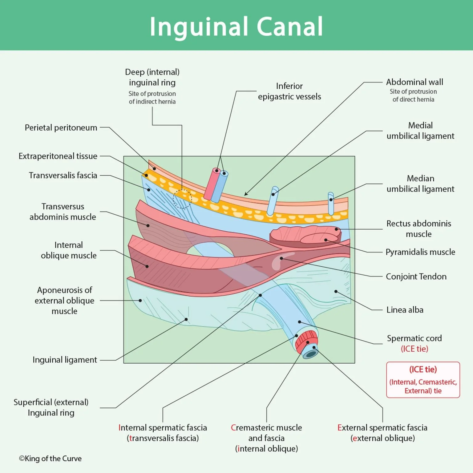
The inguinal canal runs obliquely between two openings — the deep (internal) inguinal ring and the superficial (external) inguinal ring. Its layered structure provides both passage and protection for vital anatomical contents.
| Feature | Key Points |
|---|---|
| Openings |
Deep (internal) ring — entry; site of indirect hernia. Superficial (external) ring — exit above pubic tubercle. |
| Boundaries |
|
| Contents |
Male: Spermatic cord | Female: Round ligament. Ilioinguinal nerve (travels through part of canal). |
| Fascial Coverings |
Mnemonic ICE tie:
|
| Vessel Landmark | Relation to inferior epigastric vessels: Lateral → Indirect, Medial → Direct. |
| Hernia Types |
Indirect: Through deep ring; may enter scrotum; congenital (patent processus vaginalis). Direct: Through Hesselbach’s triangle (posterior wall weakness); acquired; rarely enters scrotum. |
Contained within are several important elements, including the spermatic cord, ilioinguinal nerve, and connective tissue layers derived from the abdominal wall. To simplify the complex anatomy, students often remember the fascial coverings of the spermatic cord with the mnemonic “ICE tie”:
I – Internal spermatic fascia (from transversalis fascia)
C – Cremasteric fascia and muscle (from internal oblique)
E – External spermatic fascia (from external oblique)
⚕️ Clinical Relevance: Inguinal Hernias
The inguinal canal’s unique structure makes it vulnerable to hernias — protrusions of abdominal contents through weakened tissue. These hernias are classified based on their anatomical path:
Indirect Inguinal Hernia:
Passes through the deep inguinal ring, often extending into the scrotum.
Typically congenital, due to a patent processus vaginalis.
Located lateral to the inferior epigastric vessels.
Direct Inguinal Hernia:
Protrudes through the posterior wall of the inguinal canal, specifically Hesselbach’s triangle.
Acquired with age or increased intra-abdominal pressure.
Lies medial to the inferior epigastric vessels.
Recognizing whether a hernia is direct or indirect is vital for surgical repair, as it guides the choice of technique and prevents recurrence.
🔬 Functional and Developmental Insight
The inguinal canal also has a developmental origin, forming during fetal descent of the testes in males. The processus vaginalis, an embryonic outpouching of the peritoneum, creates the pathway through which the testes descend. Normally, this tract closes before birth — but when it doesn’t, it leaves an open channel that can lead to an indirect inguinal hernia.
In females, the canal’s function is less pronounced but still significant, as it allows passage of the round ligament that helps anchor the uterus in place.
The canal’s structural complexity — balancing mobility and support — reflects an evolutionary compromise: allowing reproductive anatomy to function while maintaining abdominal wall integrity.
💉 Diagnostic and Surgical Importance
Clinically, examination of the inguinal region helps detect hernias through palpation and imaging. Surgeons often use a combination of ultrasound and CT scanning to differentiate between direct, indirect, and femoral hernias.
During surgery, precise knowledge of the anterior and posterior walls, neurovascular structures, and fascial layers is essential to avoid complications and ensure successful outcomes.
Modern surgical techniques like laparoscopic hernia repair (TAPP and TEP approaches) rely heavily on anatomical accuracy of this region. This makes a detailed understanding of the inguinal canal not just academic — but essential for real-world medicine.
❤️ Key Takeaways
The inguinal canal is a vital structure in the lower abdomen that allows passage of reproductive and supportive tissues.
It is a common site of inguinal hernias, which are classified as direct or indirect based on their pathway.
Remembering the “ICE tie” mnemonic simplifies the fascial relationships of the canal.
Mastery of its anatomy aids in both diagnosis and surgical management.
📘 Call to Action
Want to master high-yield anatomy concepts like this one for your MCAT, USMLE, or COMLEX exams?
🧠 Dive into King of the Curve’s learning resources — where complex topics become clear through beautiful visuals, interactive quizzes, and medical storytelling.
👉 Visit King of the Curve and take your anatomy knowledge to the next level today!

Frequently Asked Questions (FAQs)
-
Aim for 4-6 focused hours, ensuring you incorporate breaks to avoid burnout.
-
Practice mindfulness techniques, take practice exams under realistic conditions, and maintain a balanced lifestyle.
-
Set short-term goals, seek support from mentors, and reward yourself for small achievements.
-
Regular exercise improves focus, reduces stress, and enhances overall mental clarity.
-
KOTC offers personalized learning tools, gamification features, and adaptive question banks to help students stay on track without burnout.