
🩺 Anatomy of the Abdominal Wall: Vessels and Muscles
Understanding the abdominal wall’s anatomy is crucial for anyone studying medicine, surgery, or human physiology. The abdominal wall is not just a protective layer; it plays a critical role in posture, internal organ support, and most importantly—vascular supply and drainage.
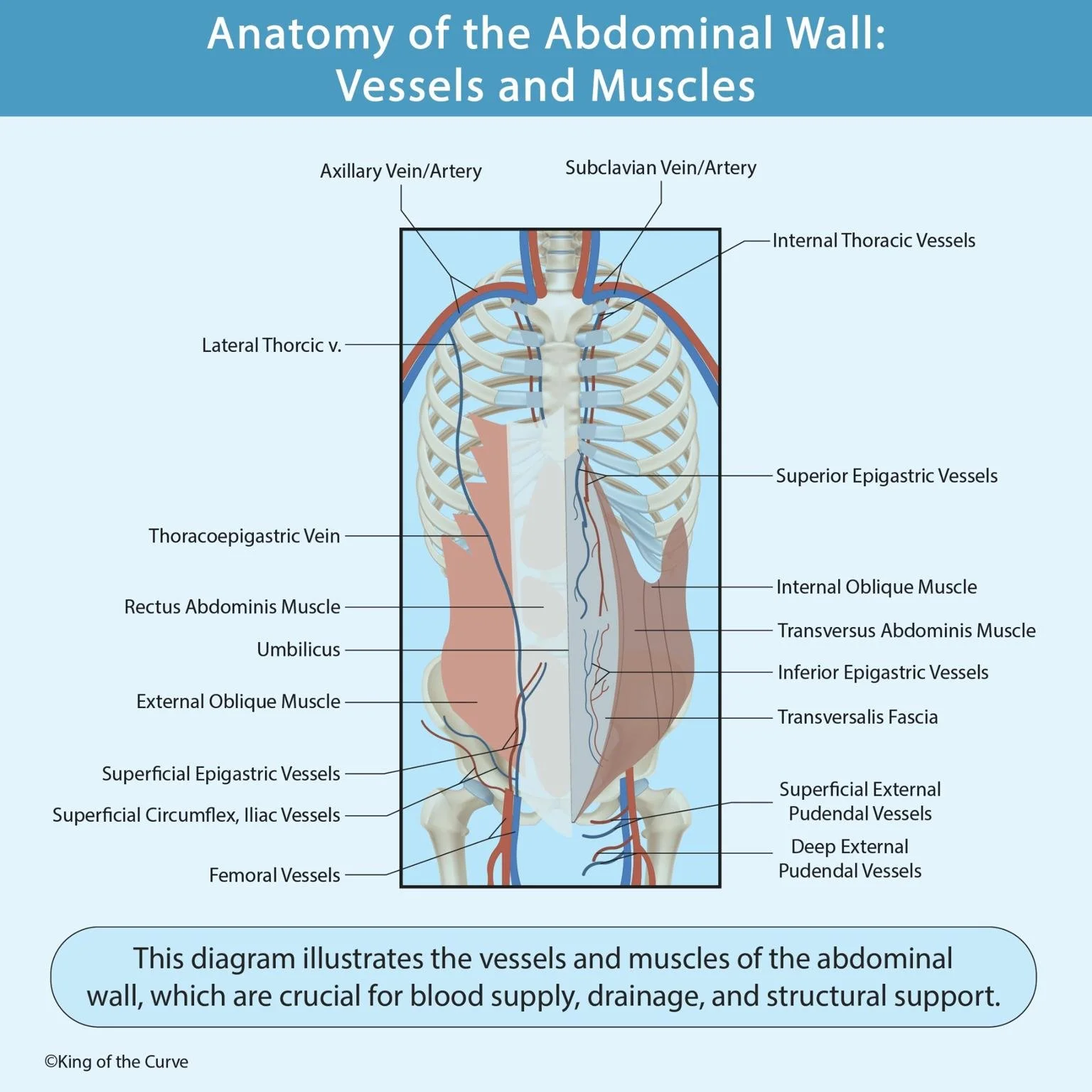
🔍 Overview of the Abdominal Wall
The abdominal wall is composed of multiple muscle layers, fascia, blood vessels, and nerves. It forms the outermost boundary of the abdominal cavity and is essential in functions like breathing, trunk movement, and protection of vital organs.
This diagram provides a comprehensive visual breakdown of the major vessels and muscles present in the abdominal wall from a frontal perspective.
🧠 Muscles of the Abdominal Wall
Muscles are color-coded and clearly labeled in this diagram:
Rectus Abdominis Muscle: Known for forming the "six-pack," it runs vertically on each side of the anterior wall and is vital for flexing the torso.
External Oblique Muscle: Located on the outer side, it allows trunk rotation and lateral flexion.
Internal Oblique Muscle: Lies beneath the external oblique and supports abdominal wall tension.
Transversus Abdominis Muscle: The deepest layer; responsible for maintaining abdominal pressure.
Transversalis Fascia: A connective tissue layer that lines the abdominal cavity and helps stabilize the core.
🩸 Key Vessels in the Abdominal Wall
Proper blood supply and venous return are critical to abdominal wall function and healing post-surgery. Highlighted vessels include:
🔼 Superior Section:
Axillary and Subclavian Veins/Arteries
Internal Thoracic Vessels
Superior Epigastric Vessels – direct continuation of the internal thoracic artery
🔽 Inferior Section:
Inferior Epigastric Vessels – branch from the external iliac artery and anastomose with superior epigastric vessels
Superficial Epigastric and Circumflex Iliac Vessels
Superficial and Deep External Pudendal Vessels
Femoral Vessels – major blood supply of the lower limb
↔️ Connecting Vessels:
Lateral Thoracic Vein
Thoracoepigastric Vein – creates an alternate venous return route
These vessels are essential during surgeries (like hernia repairs or abdominal flaps) and in conditions involving venous congestion or trauma.
🎯 Clinical Relevance
Surgical Planning: Knowing these vascular territories helps prevent accidental injury and ensures optimal incision placement.
Emergency Care: Helps in identifying internal bleeding or trauma routes.
Medical Imaging: Accurate vessel and muscle localization is key in radiological diagnosis.
📌 Summary
This anatomical chart is a concise yet detailed reference that illustrates:
✅ Muscle layers of the abdominal wall
✅ Vascular networks crucial for blood flow and drainage
✅ Clinical insights for surgical and diagnostic relevance
Understanding this structure enhances safe clinical practice and anatomical knowledge, forming a strong foundation for healthcare students and professionals alike.

Frequently Asked Questions (FAQs)
-
Aim for 4-6 focused hours, ensuring you incorporate breaks to avoid burnout.
-
Practice mindfulness techniques, take practice exams under realistic conditions, and maintain a balanced lifestyle.
-
Set short-term goals, seek support from mentors, and reward yourself for small achievements.
-
Regular exercise improves focus, reduces stress, and enhances overall mental clarity.
-
KOTC offers personalized learning tools, gamification features, and adaptive question banks to help students stay on track without burnout.