
🧬 Anatomy of a Lymph Node
Lymph nodes are small, bean-shaped structures that serve as filters for lymphatic fluid, playing a critical role in the immune system. Positioned throughout the body, especially in areas like the neck, armpits, and groin, they house immune cells that help detect and fight infections, cancers, and other foreign substances.
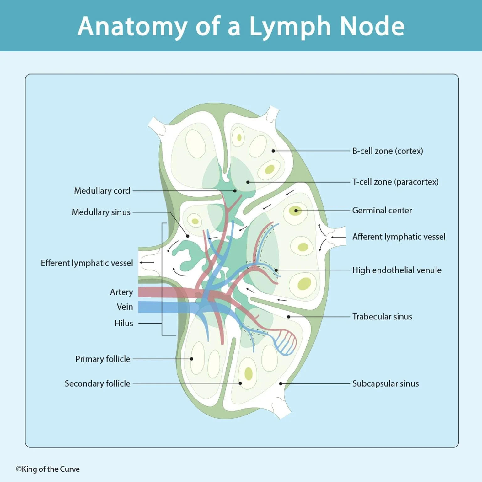
🔍 Key Structures Inside a Lymph Node
🔹 Capsule
A dense outer layer of connective tissue that encases and protects the lymph node.
🔹 Subcapsular Sinus
Located just beneath the capsule, this space collects lymph from afferent lymphatic vessels and channels it deeper into the node.
🔹 Cortex
The outer section of the lymph node, rich in B-cell zones and primary follicles, which may develop into secondary follicles with germinal centers during immune activation.
🔹 Paracortex
This inner region (also called the T-cell zone) is primarily populated by T lymphocytes and contains high endothelial venules (HEVs), specialized vessels that allow lymphocytes to enter the lymph node from the bloodstream.
🔹 Medulla
Includes:
Medullary cords: Dense clusters of lymphocytes and plasma cells.
Medullary sinuses: Pathways where lymph is filtered before exiting the node.
🧪 Immune Surveillance Pathway
Lymph enters via afferent lymphatic vessels.
It is filtered through the subcapsular, trabecular, and medullary sinuses.
Immune cells (B cells, T cells, macrophages) engage with antigens in the follicles and paracortex.
Filtered lymph exits via efferent lymphatic vessels through the hilus.
🫀 Vascular Supply
Arteries and veins enter through the hilus, providing nutrients and immune cells.
High endothelial venules (HEVs) within the paracortex allow naive lymphocytes to pass from blood into the lymph node.
🧠 Clinical Significance
Enlarged lymph nodes (lymphadenopathy) can signal infections, autoimmune conditions, or malignancies like lymphoma.
Biopsies of lymph nodes are commonly performed to diagnose systemic diseases.
Proper function is crucial for immune regulation, cancer surveillance, and vaccine response.
🩺 Quick Recap
| Structure | Function |
|---|---|
| B-cell zone (Cortex) | Hosts follicular B cells |
| T-cell zone (Paracortex) | Site of T cell activation |
| Germinal center | B cell maturation and antibody production |
| Medullary sinus | Lymph drainage path |
| Hilus | Exit site for filtered lymph and vessels |
📚 Conclusion
The lymph node is not just a passive filter—it's a dynamic immunological checkpoint. Understanding its anatomy and cellular architecture helps in diagnosing a wide range of pathologies, from infections to cancer.

Frequently Asked Questions (FAQs)
-
Aim for 4-6 focused hours, ensuring you incorporate breaks to avoid burnout.
-
Practice mindfulness techniques, take practice exams under realistic conditions, and maintain a balanced lifestyle.
-
Set short-term goals, seek support from mentors, and reward yourself for small achievements.
-
Regular exercise improves focus, reduces stress, and enhances overall mental clarity.
-
KOTC offers personalized learning tools, gamification features, and adaptive question banks to help students stay on track without burnout.