
🧠 Magnocellular vs. Parvocellular Pathways: Visual Processing Explained
The human visual system is one of the most sophisticated sensory networks in the body. Every glance, movement, and detail we perceive relies on a coordinated effort between specialized neural pathways — the magnocellular and parvocellular systems. These pathways transmit information from the retina to the brain, working together to help us see both motion and fine details with clarity.
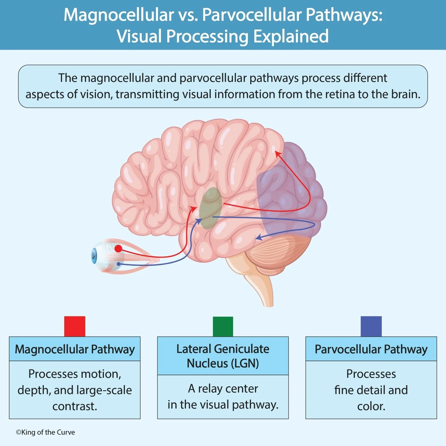
🔴 The Magnocellular Pathway: Motion and Depth Detection
The magnocellular (M) pathway processes motion, depth perception, and large-scale visual contrast. It begins in large M-type ganglion cells in the retina that transmit information quickly through thick, myelinated axons. This rapid signal transmission allows for swift responses to movement — like detecting a ball flying toward you or noticing something moving in your peripheral vision.
While it’s highly effective in detecting motion and spatial relationships, the magnocellular pathway does not process color or fine visual details. Instead, it provides a “big picture” view of dynamic changes in our environment, helping us navigate the world in real-time.
🔵 The Parvocellular Pathway: Detail and Color Recognition
In contrast, the parvocellular (P) pathway focuses on fine detail and color perception. Originating from smaller P-type ganglion cells, this pathway transmits information more slowly but with greater precision. It allows us to distinguish patterns, read text, and identify subtle differences in hues — such as the variations in a sunset or the shades of a painting.
This system works best for stationary or slowly moving objects, complementing the magnocellular pathway’s rapid motion detection. Together, they enable seamless, balanced vision — blending color, clarity, and motion into a single cohesive experience.
🟢 The Lateral Geniculate Nucleus (LGN): The Visual Relay Hub
Both the magnocellular and parvocellular pathways converge in the lateral geniculate nucleus (LGN) — a relay center within the thalamus. The LGN contains six layers: the lower two correspond to the magnocellular input, while the upper four receive parvocellular input. From there, the information travels to the primary visual cortex (V1) in the occipital lobe, where it’s integrated and interpreted.
This structured layering ensures that the brain maintains the integrity of both motion and detail signals, allowing for a complete and accurate visual representation.
📊 Comparison Table: Magnocellular vs. Parvocellular Pathways
| Feature | Magnocellular (M) Pathway | Parvocellular (P) Pathway | Lateral Geniculate Nucleus (LGN) |
|---|---|---|---|
| Primary Role | Detects motion, depth, and large-scale contrast. | Processes fine detail and color. | Relay center connecting both pathways to the visual cortex. |
| Retinal Origin | Large M-ganglion cells. | Small P-ganglion cells. | Organized into six layers (M: 1–2, P: 3–6). |
| Best At | Tracking fast movement and spatial awareness. | High-resolution detail, color, and texture. | Integrates and directs both visual signals. |
| Temporal Sensitivity | High — detects rapid changes. | Low — focused on steady images. | N/A |
| Spatial Resolution | Lower. | High. | N/A |
| Color Sensitivity | Poor. | Excellent. | Passes chromatic data to cortex. |
| Clinical Correlates | Loss → difficulty detecting motion. | Loss → impaired color and fine detail recognition. | LGN damage → visual field defects. |
| Mnemonic Tip | Think “motion and movement.” | Think “picture and pigment.” | Think “relay and refine.” |
⚡ Clinical Relevance: Understanding Visual Disorders
Lesions in the magnocellular system can lead to motion blindness or difficulty perceiving moving stimuli, while damage to the parvocellular system affects color discrimination and visual sharpness. These distinctions are crucial in diagnosing conditions like optic neuropathies, glaucoma, and certain cortical vision deficits.
Understanding these pathways also informs how visual processing disorders manifest — helping clinicians interpret symptoms like blurring, delayed tracking, or motion perception issues.
🚀 Call to Action
At King of the Curve, we believe that complex neuroscience doesn’t have to be intimidating. By combining visuals and concise explanations, we make medical concepts easier to grasp and remember.
💡 Continue exploring our visual learning library for more detailed diagrams, high-yield insights, and interactive anatomy content.

Frequently Asked Questions (FAQs)
-
Aim for 4-6 focused hours, ensuring you incorporate breaks to avoid burnout.
-
Practice mindfulness techniques, take practice exams under realistic conditions, and maintain a balanced lifestyle.
-
Set short-term goals, seek support from mentors, and reward yourself for small achievements.
-
Regular exercise improves focus, reduces stress, and enhances overall mental clarity.
-
KOTC offers personalized learning tools, gamification features, and adaptive question banks to help students stay on track without burnout.