
🩺 Understanding the Segments of the Duodenum
The duodenum is the first section of the small intestine and serves as the meeting point for food from the stomach, bile from the gallbladder, and enzymes from the pancreas. It’s a key player in digestion and nutrient breakdown. The duodenum is divided into four parts, each with distinct anatomy, blood supply, and clinical importance.
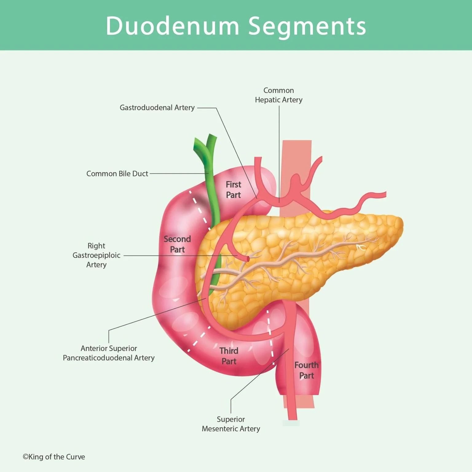
1️⃣ First Part (Superior Part)
📍 Location: Begins at the pylorus of the stomach and curves towards the right.
⚙️ Function: Receives chyme from the stomach and begins mixing with bile and enzymes.
🩸 Blood Supply: Mainly from the gastroduodenal artery and branches like the right gastroepiploic artery.
💡 Clinical Note: Most common site for duodenal ulcers due to acid exposure.
2️⃣ Second Part (Descending Part)
📍 Location: Runs vertically along the right side of the vertebral column.
⚙️ Function: Serves as the entry point for bile and pancreatic secretions via the major duodenal papilla.
🩸 Blood Supply: Supplied by both the gastroduodenal artery and superior mesenteric artery branches.
💡 Clinical Note: Important in diagnosing biliary obstructions.
3️⃣ Third Part (Horizontal Part)
📍 Location: Crosses from right to left over the inferior vena cava and aorta, under the superior mesenteric artery.
⚙️ Function: Moves digested contents towards the ascending duodenum.
🩸 Blood Supply: Primarily from the superior mesenteric artery.
💡 Clinical Note: Can be compressed between the SMA and aorta, leading to Superior Mesenteric Artery Syndrome.
4️⃣ Fourth Part (Ascending Part)
📍 Location: Runs upward to join the jejunum at the duodenojejunal flexure.
⚙️ Function: Passes contents to the jejunum for nutrient absorption.
🩸 Blood Supply: Branches from the superior mesenteric artery.
💡 Clinical Note: Supported by the ligament of Treitz, a key landmark in GI surgery.
📊 Table: Duodenum Segments Overview
| Segment | Location | Function | Blood Supply | Clinical Significance |
|---|---|---|---|---|
| First Part | After pylorus; curves right | Mixes chyme with bile & enzymes | Gastroduodenal, Right gastroepiploic | Common site of ulcers |
| Second Part | Right side of spine (descending) | Bile & pancreatic entry (major papilla) | Gastroduodenal + SMA | Biliary obstruction focus |
| Third Part | Horizontal; under SMA | Transit toward 4th part | SMA branches | SMA compression syndrome risk |
| Fourth Part | Ascends to jejunum | Passes contents to jejunum | SMA branches | Ligament of Treitz landmark |
💬 Final Thoughts
Knowing the anatomy and vascular supply of each duodenal segment is crucial for clinicians, surgeons, and medical students. It helps in diagnosing conditions like ulcers, obstructions, and vascular compression syndromes, and in performing safe abdominal surgeries.

Frequently Asked Questions (FAQs)
-
Aim for 4-6 focused hours, ensuring you incorporate breaks to avoid burnout.
-
Practice mindfulness techniques, take practice exams under realistic conditions, and maintain a balanced lifestyle.
-
Set short-term goals, seek support from mentors, and reward yourself for small achievements.
-
Regular exercise improves focus, reduces stress, and enhances overall mental clarity.
-
KOTC offers personalized learning tools, gamification features, and adaptive question banks to help students stay on track without burnout.