
🧠 Introduction to Cranial Nerve Exits and Pathways
The human brain is an extraordinary organ that communicates with the rest of the body through a network of twelve cranial nerves. These nerves pass through specific openings in the skull known as foramina or canals. Understanding their exits and pathways is crucial for medical students, neurologists, and surgeons, as it helps in diagnosing and managing neurological disorders.
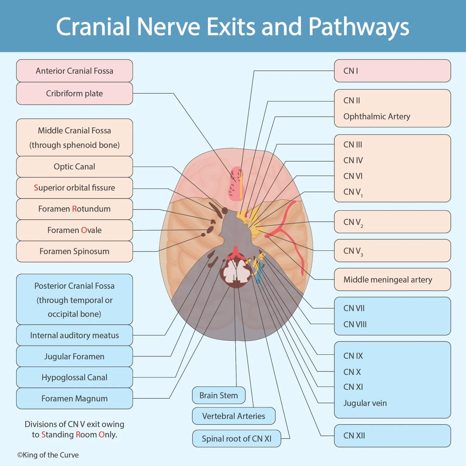
📍 The Anterior Cranial Fossa and Its Nerve Exits
The anterior cranial fossa houses the frontal lobes of the brain and is associated primarily with the cribriform plate of the ethmoid bone. This structure allows the Olfactory Nerve (CN I) to pass through, enabling our sense of smell. Damage to this area can lead to anosmia (loss of smell), which may be caused by trauma or infection.
🔍 The Middle Cranial Fossa Pathways
The middle cranial fossa contains several important openings through the sphenoid bone. These include:
Optic Canal: Transmits the Optic Nerve (CN II) and the Ophthalmic Artery.
Superior Orbital Fissure: Allows passage of CN III, CN IV, CN VI, and CN V₁, controlling most eye movements.
Foramen Rotundum: Transmits CN V₂.
Foramen Ovale: Carries CN V₃.
Foramen Spinosum: Contains the middle meningeal artery, important for blood supply to the meninges.
🩺 The Posterior Cranial Fossa and Nerve Functions
The posterior cranial fossa houses critical structures of the brainstem and cerebellum. Key openings include:
Internal Auditory Meatus: Transmits CN VII (Facial) and CN VIII (Vestibulocochlear).
Jugular Foramen: Carries CN IX, CN X, CN XI, and the jugular vein.
Hypoglossal Canal: Contains CN XII.
Foramen Magnum: Allows the passage of the spinal cord, vertebral arteries, and the spinal root of CN XI.
📊 Summary Table of Cranial Nerve Exits
Cranial Nerve Exits and Pathways Table
| Foramen / Canal | Cranial Nerve(s) & Structures |
|---|---|
| Cribriform Plate | CN I |
| Optic Canal | CN II, Ophthalmic Artery |
| Superior Orbital Fissure | CN III, CN IV, CN VI, CN V1 |
| Foramen Rotundum | CN V2 |
| Foramen Ovale | CN V3 |
| Foramen Spinosum | Middle meningeal artery |
| Internal Auditory Meatus | CN VII, CN VIII |
| Jugular Foramen | CN IX, CN X, CN XI, Jugular vein |
| Hypoglossal Canal | CN XII |
| Foramen Magnum | Spinal cord, Vertebral arteries, Spinal root of CN XI |
📢 Call to Action
Mastering cranial nerve exits and pathways is a vital step in becoming confident in neuroanatomy. If you want to strengthen your clinical knowledge and ace your exams, keep practicing with detailed anatomical diagrams and mnemonic tools. Explore more interactive neuroanatomy resources with us and elevate your learning!

Frequently Asked Questions (FAQs)
-
Aim for 4-6 focused hours, ensuring you incorporate breaks to avoid burnout.
-
Practice mindfulness techniques, take practice exams under realistic conditions, and maintain a balanced lifestyle.
-
Set short-term goals, seek support from mentors, and reward yourself for small achievements.
-
Regular exercise improves focus, reduces stress, and enhances overall mental clarity.
-
KOTC offers personalized learning tools, gamification features, and adaptive question banks to help students stay on track without burnout.